
Page 27 - Cardiac Nursing
P. 27
92806_c01.qxd 11/21/11 10:30 AM Page 3
CHAPTER 1 / Cardiac Anatomy and Physiology 3
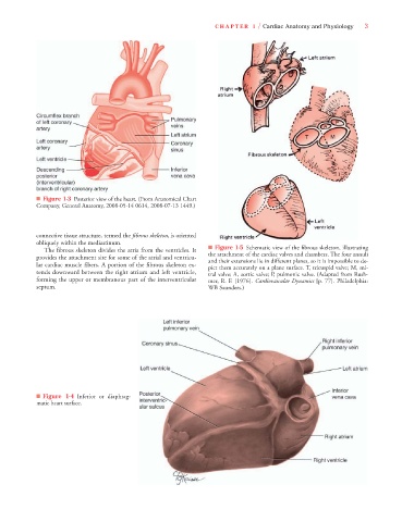
■ Figure 1-3 Posterior view of the heart. (From Anatomical Chart
Company, General Anatomy, 2008-05-14 0614, 2008-07-13 1449.)
connective tissue structure, termed the fibrous skeleton, is oriented
obliquely within the mediastinum.
The fibrous skeleton divides the atria from the ventricles. It ■ Figure 1-5 Schematic view of the fibrous skeleton, illustrating
provides the attachment site for some of the atrial and ventricu- the attachment of the cardiac valves and chambers. The four annuli
and their extensions lie in different planes, so it is impossible to de-
lar cardiac muscle fibers. A portion of the fibrous skeleton ex- pict them accurately on a plane surface. T, tricuspid valve; M, mi-
tends downward between the right atrium and left ventricle, tral valve; A, aortic valve; P, pulmonic valve. (Adapted from Rush-
forming the upper or membranous part of the interventricular mer, R. F. [1976]. Cardiovascular Dynamics [p. 77]. Philadelphia:
septum. WB Saunders.)
■ Figure 1-4 Inferior or diaphrag-
matic heart surface.