
Page 267 - Cardiac Nursing
P. 267
1
1
009
009
0:4
7 A
M
0:4
7 A
xd
3
q
xd
3
6/2
6/2
0/0
0/0
M
p
p
A
A
p
ara
ara
t
t
g
g
Pa
Pa
g
43
43
e 2
e 2
LWB
K34
p
p
K34
10_
10_
0-c
0-c
21
44.
44.
q
q
1-2
LWB
21
LWBK340-c10_ pp211-244.qxd 30/06/2009 10:47 AM Page 243 Aptara
1-2
C HAPTER 1 0 / History Taking and Physical Examination 243
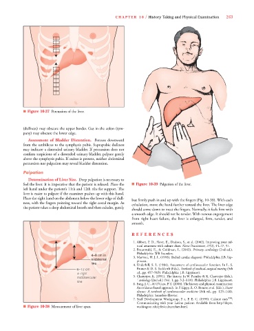
■ Figure 10-37 Percussion of the liver.
(dullness) may obscure the upper border. Gas in the colon (tym-
pany) may obscure the lower edge.
Assessment of Bladder Distention. Percuss downward
from the umbilicus to the symphysis pubis. Suprapubic dullness
may indicate a distended urinary bladder. If percussion does not
confirm suspicions of a distended urinary bladder, palpate gently
above the symphysis pubis. If ascites is present, neither abdominal
percussion nor palpation may reveal bladder distention.
Palpation
Determination of Liver Size. Deep palpation is necessary to
feel the liver. It is imperative that the patient is relaxed. Place the ■ Figure 10-39 Palpation of the liver.
left hand under the patient’s 11th and 12th ribs for support. The
liver is easier to palpate if the examiner pushes up with this hand.
Place the right hand on the abdomen below the lower edge of dull- but firmly push in and up with the fingers (Fig. 10-39). With each
ness, with the fingers pointing toward the right costal margin. As exhalation, move the hand further toward the liver. The liver edge
the patient takes a deep abdominal breath and then exhales, gently
should come down to meet the fingers. Normally, it feels firm with
a smooth edge. It should not be tender. With venous engorgement
from right heart failure, the liver is enlarged, firm, tender, and
smooth.
REFE R E NC ES
1. Abbott, P. D., Short, E., Dodson, S., et al. (2002). Improving your cul-
7
7
tural awareness with culture clues. Nurse Practitioner, 27(2), 44–47, 51.
2. Braunwald, E., & Goldman, L. (2002). Primary cardiology (2nd ed.).
Philadelphia: WB Saunders.
4– 8 8 8 8c c c c c c c cm i i i in n n n n n n 3. Marriott, H. J. L. (1993). Bedside cardiac diagnosis. Philadelphia: J.B. Lip-
8
8
8
c
m
m
cm
cm
c
–8
–8
–8
4–8
4–8 cm in
4 4 4 4 4–
4–8
–
4–
8c
8
4–
–
–
–
m
m
m
m
in
m
m
m
m
in
in
in
m
m
m
m
m
midste
rn
r
e
er
e
e
r
r
e
midst te er rn pincott.
n
n
na
n
n
na
rn
n
n
n
s
st
st
er
e
s
st
te
te
te
s
st
st
ds
er
d
d
idsternanal
i i id
e
d
ds
d
ds
d
line
l l l l line 4. Underhill, S. L. (1984). Assessment of cardiovascular function. In L. S.
m
m
m
m
m
m
m
m
m
m
m
m
2
2
2
2
1
1
2
2
c
c
2
2
2
2
1
–
–
–
–
6–12 cm Bruner & D. S. Suddarth (Eds.), Textbook of medical–surgical nursing (5th
6 6 6 6 6 6 6 6 6–
–
–
1
1
–
–
–
–
c
c
c
c
c
c
c
g
g
g
gh
gh
n r
n r
n r
gh
g
ri
ri
ri
ig
ri
gh
gh
gh
ri
ri
n r
h
in
ht
ht
ht
ht
in r
ht
ht
in right
i i in in in in in in in in in ri i ig g g gh t t t ed., pp. 457–563). Philadelphia: J.B. Lippincott.
in
in r
vi
vi
dc
mid
ic
idc
mid cla vic cu la a ar r r r r r 5. Chatterjee, K. (1991). The history. In W. Parmley & K. Chatterjee (Eds.),
midc
vic
mid
midclavicular
mid
mid
mid
m m mi
vi
vic
v
mi
midc
midc
av
av
av
av
av
a
a
a
av
ar
la
cla
ar
cla
cla
cla
la
la
la
ar
cla
cu
cu
cu
cu
cu
dc
dcl
dcl
ic
dc
c
a
u
u
av
u
ul
c
ul
ul
ula
line
line
line
line
ne
ne
ine
lin
line
l li li lin e e e e Cardiology (2nd ed.) (Vol. 1, pp. 3.2–3.10). Philadelphia: J.B. Lippincott.
line
line
line
line
6.Fang, J. C., & O’Gara, P. T. (2008). The history and physical examination:
An evidence-based approach. In P. Lippy, R. O. Bonow, et al. (Eds.), Heart
disease: A textbook of cardiovascular medicine (8th ed., pp. 125–148).
Philadelphia: Saunders-Elsevier.
7. Staff Development Workgroup, P. a. F. E. C. (1999). Culture cues TM :
Communicating with your Latino patient: Available from http//depts.
■ Figure 10-38 Measurement of liver span. washington.edu/pfes/cultureclues.html.