
Page 264 - Cardiac Nursing
P. 264
6/2
009
6/2
0/0
0/0
0:4
0:4
1
009
1
3
44.
q
44.
1-2
1-2
xd
3
xd
q
q
7 A
p
p
A
40
A
ara
ara
t
p
t
40
Pa
Pa
M
7 A
M
e 2
e 2
g
g
g
LWBK340-c10_ p p pp211-244.qxd 30/06/2009 10:47 AM Page 240 Aptara
K34
21
21
K34
10_
10_
0-c
0-c
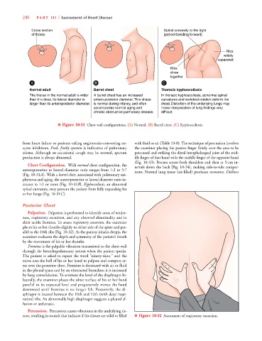
240 P A R T III / Assessment of Heart Disease
Cross section Spinal convexity to the right
of thorax (patient bending forward)
Ribs
widely
se parated
Ribs
close
together
A B C
Normal adult Barrel chest Thoracic kyphoscoliosis
The thorax in the normal adult is wider A barrel chest has an increased In thoracic kyphoscoliosis, abnormal spinal
than it is deep. Its lateral diameter is antero-posterior diameter. This shape curvatures and vertebral rotation deform the
larger than its anteroposterior diameter. is normal during infancy, and often chest. Distortion of the underlying lungs may
accompanies normal aging and make interpretation of lung findings very
chronic obstructive pulmonary disease. difficult.
■ Figure 10-31 Chest wall configurations. (A) Normal. (B) Barrel chest. (C) Kyphoscoliosis.
from heart failure or patients taking angiotensin-converting en- with fluid or air (Table 10-8). The technique of percussion involves
zyme inhibitors. Pink, frothy sputum is indicative of pulmonary the examiner placing the passive finger firmly over the area to be
edema. Although an occasional cough may be normal, sputum percussed and striking the distal interphalangeal joint of the mid-
production is always abnormal. dle finger of that hand with the middle finger of the opposite hand
(Fig. 10-33). Percuss across both shoulders and then at 5-cm in-
Chest Configuration. With normal chest configuration, the tervals down the back (Fig. 10-34), making side-to-side compar-
anteroposterior to lateral diameter ratio ranges from 1:2 to 5:7 isons. Normal lung tissue (air-filled) produces resonance. Dullness
t
(Fig. 10-31A). With a barrel chest, associated with pulmonary em-
t
physema and aging, the anteroposterior to lateral diameter ratio in-
creases to 1:1 or more (Fig. 10-31B). Kyphoscoliosis, an abnormal
B
spinal curvature, may prevent the patient from fully expanding his
or her lungs (Fig. 10-31C).
Posterior Chest
Palpation. Palpation is performed to identify areas of tender-
ness, respiratory excursion, and any observed abnormality and to
elicit tactile fremitus. To assess respiratory excursion, the examiner
places his or her thumbs slightly to either side of the spine and par-
allel to the 10th ribs (Fig. 10-32). As the patient inhales deeply, the
examiner evaluates the depth and symmetry of the patient’s breath
by the movement of his or her thumbs.
Fremitus is the palpable vibration transmitted to the chest wall
through the bronchopulmonary system when the patient speaks.
The patient is asked to repeat the word “ninety-nine,” and the
nurse uses the ball of his or her hand to palpate and compare ar-
eas over the posterior chest. Fremitus is decreased with air or fluid
in the pleural space and by an obstructed bronchus; it is increased
by lung consolidation. To estimate the level of the diaphragm bi-
laterally, the examiner places the ulnar surface of his or her hand
parallel to its expected level and progressively moves the hand
downward until fremitus is no longer felt. Posteriorly, the di-
aphragm is located between the 10th and 12th (with deep inspi-
ration) ribs. An abnormally high diaphragm suggests a pleural ef-
fusion or atelectasis.
Percussion. Percussion causes vibrations in the underlying tis-
sues, resulting in sounds that indicate if the tissues are solid or filled ■ Figure 10-32 Assessment of respiratory excursion.