
Page 263 - Cardiac Nursing
P. 263
009
009
6/2
0/0
6/2
0:4
7 A
0:4
1
1
0/0
q
q
44.
1-2
44.
3
3
xd
q
xd
p
p
A
39
A
ara
ara
t
p
t
39
Pa
Pa
M
7 A
M
e 2
e 2
g
g
g
LWBK340-c10_
21
K34
10_
0-c
LWB K34 0-c 10_ p p pp211-244.qxd 30/06/2009 10:47 AM Page 239 Aptara
1-2
21
LWB
C HAPTER 1 0 / History Taking and Physical Examination 239
subaortic stenosis and mitral valve prolapse. Sudden standing has tory manifestations seen in patients with heart disease. The room
the opposite effect; it reduces venous return and causes most should be quiet and the patient’s chest exposed. Proceed in a sys-
murmurs, except hypertrophic cardiomyopathy and mitral valve tematic manner: inspect, palpate, percuss, and auscultate. Always
prolapse, to decrease. Squatting simultaneously increases venous compare one side with the other; always place the stethoscope in
return and systemic vascular resistance. 6 direct contact with the chest wall. Begin with examination of the
Postextrasystolic beats, if followed by a pause, increase ventricular posterior chest, if possible, with the patient sitting upright and
filling and cardiac contractility. Similar hemodynamic changes oc- arms folded across the chest. Follow with assessment of the ante-
cur with diastolic pauses in atrial fibrillation and sinus arrhythmia. 6 rior chest with the patient lying down. Only the upper and lower
Isometric exercise increases systemic vascular resistance, arterial lobes of the lung are accessible by posterior chest examination; to
pressure, heart rate, cardiac output, left ventricular filling pressure, assess the right middle lobe, the lateral and anterior chest must be
and heart size. Using a calibrated handgrip device, the patient sus- examined (Fig. 10-29). 8
tains the handgrip for 20 to 30 seconds. The handgrip enhances
S 3 and S 4 and aortic regurgitant murmurs. Avoid isometric exer- Inspection
cise in patients with myocardial ischemia or ventricular arrhyth-
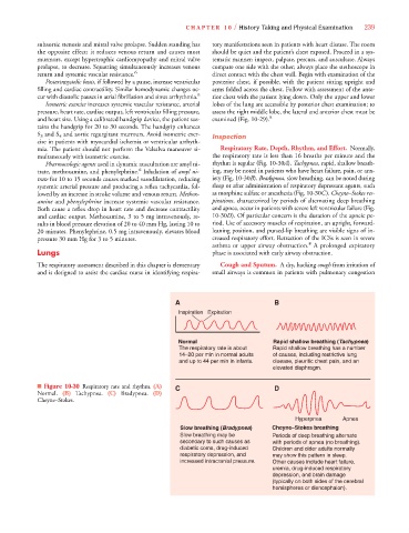
mia. The patient should not perform the Valsalva maneuver si- Respiratory Rate, Depth, Rhythm, and Effort. Normally,
multaneously with isometric exercise. the respiratory rate is less than 16 breaths per minute and the
0
0
Pharmacologic agents used in dynamic auscultation are amyl ni- rhythm is regular (Fig. 10-30A). Tachypnea, rapid, shallow breath-
6
trate, methoxamine, and phenylephrine. Inhalation of amyl ni- ing, may be noted in patients who have heart failure, pain, or anx-
trate for 10 to 15 seconds causes marked vasodilatation, reducing iety (Fig. 10-30B).B Bradypnea, slow breathing, can be noted during
systemic arterial pressure and producing a reflex tachycardia, fol- sleep or after administration of respiratory depressant agents, such
lowed by an increase in stroke volume and venous return. Methox- as morphine sulfate or anesthesia (Fig. 10-30C). Cheyne–Stokes res-
amine and phenylephrine increase systemic vascular resistance. pirations, characterized by periods of alternating deep breathing
Both cause a reflex drop in heart rate and decrease contractility and apnea, occur in patients with severe left ventricular failure (Fig.
and cardiac output. Methoxamine, 3 to 5 mg intravenously, re- 10-30D). Of particular concern is the duration of the apneic pe-
sults in blood pressure elevation of 20 to 40 mm Hg, lasting 10 to riod. Use of accessory muscles of respiration, an upright, forward-
20 minutes. Phenylephrine, 0.5 mg intravenously, elevates blood leaning position, and pursed-lip breathing are visible signs of in-
pressure 30 mm Hg for 3 to 5 minutes. creased respiratory effort. Retraction of the ICSs is seen in severe
8
asthma or upper airway obstruction. A prolonged expiratory
Lungs phase is associated with early airway obstruction.
The respiratory assessment described in this chapter is elementary Cough and Sputum. A dry, hacking cough from irritation of
and is designed to assist the cardiac nurse in identifying respira- small airways is common in patients with pulmonary congestion
A B
Inspiration Expiration
a
Normal Rapid shallow breathing (Tachypnea)
a
The respiratory rate is about Rapid shallow breathing has a number
14–20 per min in normal adults of causes, including restrictive lung
and up to 44 per min in infants. disease, pleuritic chest pain, and an
elevated diaphragm.
■ Figure 10-30 Respiratory rate and rhythm. (A) C D
Normal. (B) Tachypnea. (C) Bradypnea. (D)
Cheyne–Stokes.
Hyperpnea Apnea
a
Slow breathing (Bradypnea) Cheyne–Stokes breathing
a
Slow breathing may be Periods of deep breathing alternate
secondary to such causes as with periods of apnea (no breathing).
diabetic coma, drug-induced Children and older adults normally
respiratory depression, and may show this pattern in sleep.
increased intracranial pressure. Other causes include heart failure,
uremia, drug-induced respiratory
depression, and brain damage
(typically on both sides of the cerebral
hemispheres or diencephalon).