
Page 319 - Cardiac Nursing
P. 319
Pa
g
M
Pa
g
e 2
95
g
e 2
1
1
/09
/09
0:2
7 P
M
0:2
7 P
95
a
a
ara
a
a
c.
c.
In
In
p
p
A
A
p
ara
ara
t
t
1-2
1-2
LWB K34 0-c 14_ p p pp291-299.qxd 6/29/09 10:27 PM Page 295 Aptara Inc.
99.
q
q
99.
K34
0-c
14_
LWBK340-c14_
29
29
LWB
6
6
/29
/29
6
q
xd
xd
C HAPTER 1 4 / Nuclear, Magnetic Resonance, and Computed Tomography Imaging 295
stenoses have been performed in research studies, MRI is not
commonly performed clinically for that purpose. However, MRI
allows descriptions of anomalous coronary artery origins and
courses of the arteries near the great vessels. The pericardium can
be visualized by MRI, and thus descriptions of pericardial thick-
ness and locations of pericardial effusions can be provided. Ven-
tricular volumes and function can be obtained by MRI because of
the excellent visualization of endocardial borders. 28–31 The right
ventricle is seen in limited views with echo and most commonly,
the function is described qualitatively. As opposed to echo, quan-
titation of RV function can be performed in terms of RV volumes
and EFs. Trans-valvular velocities, trans-valvular pressure gradi-
ents, and valve areas are all measures of the severity of valvular
stenosis. 32–35 Typically, these measures are obtained by echo but
can also be evaluated by MRI. Quantitation of the amount
of valvular regurgitation (regurgitant volumes) can also be
performed. 36,37
Myocardial perfusion can be used to assess the presence or ab-
sence of flow-limited coronary artery disease. 38,39 Exercise testing
is not performed. Instead, perfusion at stress can be assessed with
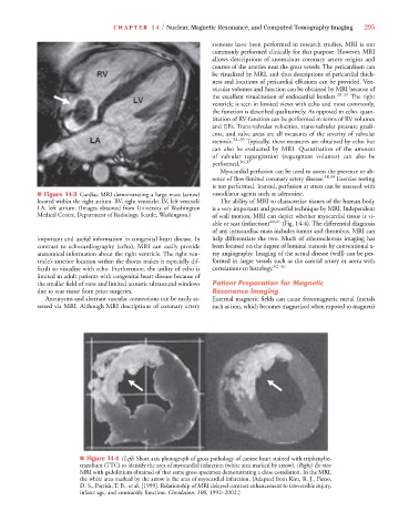
■ Figure 14-3 Cardiac MRI demonstrating a large mass (arrow) vasodilator agents such as adenosine.
located within the right atrium. RV, right ventricle; LV, left ventricle The ability of MRI to characterize tissues of the human body
LA, left atrium. (Images obtained from University of Washington is a very important and powerful technique by MRI. Independent
Medical Center, Department of Radiology, Seattle, Washington.) of wall motion, MRI can depict whether myocardial tissue is vi-
able or scar (infarction) 40,41 (Fig. 14-4). The differential diagnosis
of any intracardiac mass includes tumor and thrombus. MRI can
important and useful information in congenital heart disease. In help differentiate the two. Much of atherosclerosis imaging has
contrast to echocardiography (echo), MRI can easily provide been focused on the degree of luminal stenosis by conventional x-
anatomical information about the right ventricle. The right ven- ray angiography. Imaging of the actual disease (wall) can be per-
tricle’s anterior location within the thorax makes it especially dif- formed in larger vessels such as the carotid artery or aorta with
ficult to visualize with echo. Furthermore, the utility of echo is correlations to histology. 42–46
limited in adult patients with congenital heart disease because of
the smaller field of view and limited acoustic ultrasound windows Patient Preparation for Magnetic
due to scar tissue from prior surgeries. Resonance Imaging
Aneurysms and aberrant vascular connections can be easily as- External magnetic fields can cause ferromagnetic metal (metals
sessed via MRI. Although MRI descriptions of coronary artery such as iron, which becomes magnetized when exposed to magnets)
■ Figure 14-4 (Left) Short axis photograph of gross pathology of canine heart stained with triphenylte-
trazolium (TTC) to identify the area of myocardial infarction (white area marked by arrow). (Right) Ex vivo
MRI with gadolinium obtained of that same gross specimen demonstrating a close correlation. In the MRI,
the white area marked by the arrow is the area of myocardial infarction. (Adapted from Kim, R. J., Fieno,
D. S., Parrish, T. B., et al. [1999]. Relationship of MRI delayed contrast enhancement to irreversible injury,
infarct age, and contractile function. Circulation, 100, 1992–2002.)