
Page 321 - Cardiac Nursing
P. 321
1
1
0:2
/29
/09
/09
M
M
Pa
0:2
7 P
7 P
/29
99.
q
q
1-2
1-2
99.
6
6
6
q
xd
xd
Pa
ara
a
a
t
ara
ara
In
c.
c.
a
a
In
t
e 2
e 2
97
g
g
g
p
p
p
97
A
A
14_
0-c
29
29
LWB
LWB K34 0-c 14_ p p pp291-299.qxd 6/29/09 10:27 PM Page 297 Aptara Inc.
K34
LWBK340-c14_
CHAP TE R 1 4 / Nuclear, Magnetic Resonance, and Computed Tomography Imaging 297
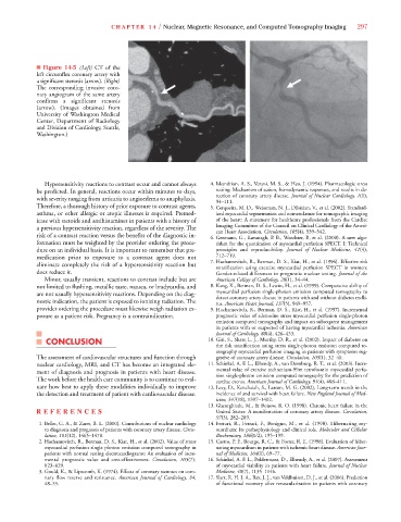
■ Figure 14-5 (Left) CT of the
left circumflex coronary artery with
a significant stenosis (arrow). (Right)
The corresponding invasive coro-
nary angiogram of the same artery
confirms a significant stenosis
(arrow). (Images obtained from
University of Washington Medical
Center, Department of Radiology
and Division of Cardiology, Seattle,
Washington.)
Hypersensitivity reactions to contrast occur and cannot always 4. Iskandrian, A. S., Verani, M. S., & Heo, J. (1994). Pharmacologic stress
be predicted. In general, reactions occur within minutes to days, testing: Mechanism of action, hemodynamic responses, and results in de-
with severity ranging from urticaria to angioedema to anaphylaxis. tection of coronary artery disease. Journal of Nuclear Cardiology, 1(1),
94–111.
Therefore, a thorough history of prior exposure to contrast agents, 5. Cerqueira, M. D., Weissman, N. J., Dilsizian, V., et al. (2002). Standard-
asthma, or other allergic or atopic illnesses is required. Premed- ized myocardial segmentation and nomenclature for tomographic imaging
icate with steroids and antihistamines in patients with a history of of the heart: A statement for healthcare professionals from the Cardiac
a previous hypersensitivity reaction, regardless of the severity. The Imaging Committee of the Council on Clinical Cardiology of the Ameri-
can Heart Association. Circulation, 105(4), 539–542.
risk of a contrast reaction versus the benefits of the diagnostic in- 6. Germano, G., Kavanagh, P. B., Waechter, P., et al. (2000). A new algo-
formation must be weighted by the provider ordering the proce- rithm for the quantitation of myocardial perfusion SPECT. I: Technical
dure on an individual basis. It is important to remember that pre- principles and reproducibility. Journal of Nuclear Medicine, 41(4),
medication prior to exposure to a contrast agent does not 712–719.
eliminate completely the risk of a hypersensitivity reaction but 7. Hachamovitch, R., Berman, D. S., Kiat, H., et al. (1996). Effective risk
stratification using exercise myocardial perfusion SPECT in women:
does reduce it. Gender-related differences in prognostic nuclear testing. Journal of the
Minor, usually transient, reactions to contrast include but are American College of Cardiology, 28(1), 34–44.
not limited to flushing, metallic taste, nausea, or bradycardia, and 8. Kang, X., Berman, D. S., Lewin, H., et al. (1999). Comparative ability of
are not usually hypersensitivity reactions. Depending on the diag- myocardial perfusion single-photon emission computed tomography to
detect coronary artery disease in patients with and without diabetes melli-
nostic indication, the patient is exposed to ionizing radiation. The tus. American Heart Journal, 137(5), 949–957.
7
7
provider ordering the procedure must likewise weigh radiation ex- 9. Hachamovitch, R., Berman, D. S., Kiat, H., et al. (1997). Incremental
posure as a patient risk. Pregnancy is a contraindication. prognostic value of adenosine stress myocardial perfusion single-photon
emission computed tomography and impact on subsequent management
in patients with or suspected of having myocardial ischemia. American
Journal of Cardiology, 80(4), 426–433.
CONCLUSION 10. Giri, S., Shaw, L. J., Murthy, D. R., et al. (2002). Impact of diabetes on
the risk stratification using stress single-photon emission computed to-
mography myocardial perfusion imaging in patients with symptoms sug-
The assessment of cardiovascular structures and function through gestive of coronary artery disease. Circulation, 105(1), 32–40.
nuclear cardiology, MRI, and CT has become an integrated ele- 11. Schinkel, A. F. L., Elhendy, A., van Domburg, R. T., et al. (2003). Incre-
ment of diagnosis and prognosis in patients with heart disease. mental value of exercise technetium-99m tetrofosmin myocardial perfu-
sion single-photon emission computed tomography for the prediction of
The work before the health care community is to continue to eval- cardiac events. American Journal of Cardiology, 91(4), 408–411.
uate how best to apply these modalities individually to improve 12. Levy, D., Kenchaiah, S., Larson, M. G. (2002). Long-term trends in the
the detection and treatment of patient with cardiovascular disease. incidence of and survival with heart failure. New England Journal of Med-
7
icine, 347(18), 1397–1402.
7
13. Gheorghiade, M., & Bonow, R. O. (1998). Chronic heart failure in the
REFEREN C E S United States: A manifestation of coronary artery disease. Circulation,
97(3), 282–289.
7
7
1. Beller, G. A., & Zaret, B. L. (2000). Contributions of nuclear cardiology 14. Ferrari, R., Ferrari, F., Benigno, M., et al. (1998). Hibernating my-
to diagnosis and prognosis of patients with coronary artery disease. Circu- ocardium: Its pathophysiology and clinical role. Molecular and Cellular
lation, 101(12), 1465–1478. Biochemistry, 186(1/2), 195–199.
6
6
2. Hachamovitch, R., Berman, D. S., Kiat, H., et al. (2002). Value of stress 15. Castro, P. F., Bourge, R. C., & Foster, R. E. (1998). Evaluation of hiber-
myocardial perfusion single photon emission computed tomography in nating myocardium in patients with ischemic heart disease. American Jour-
patients with normal resting electrocardiograms: An evaluation of incre- nal of Medicine, 104(1), 69–77.
mental prognostic value and cost-effectiveness. Circulation, 105(7), 16. Schinkel, A. F. L., Poldermans, D., Elhendy, A., et al. (2007). Assessment
823–829. of myocardial viability in patients with heart failure. Journal of Nuclear
3. Gould, K., & Lipscomb, K. (1974). Effects of coronary stenoses on coro- Medicine, 48(7), 1135–1146.
nary flow reserve and resistance. American Journal of Cardiology, 34, 17. Slart, R. H. J. A., Bax, J. J., van Veldhuisen, D. J., et al. (2006). Prediction
48–55. of functional recovery after revascularization in patients with coronary