
Page 316 - Cardiac Nursing
P. 316
LWBK340-c14_p291-299.qxd 6/29/09 10:27 PM Page 292 Aptara Inc.
292 PA R T III / Assessment of Heart Disease
appropriately dilate in response to increasing myocardial work re-
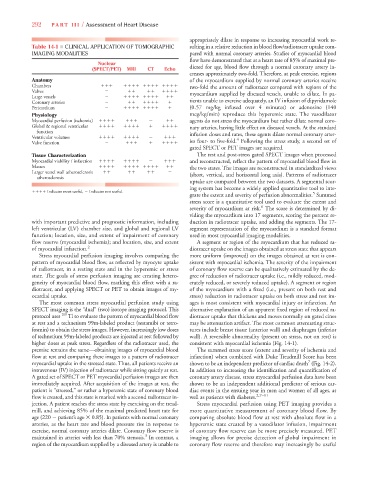
Table 14-1 ■ CLINICAL APPLICATION OF TOMOGRAPHIC sulting in a relative reduction in blood flow/radiotracer uptake com-
IMAGING MODALITIES pared with normal coronary arteries. Studies of myocardial blood
flow have demonstrated that at a heart rate of 85% of maximal pre-
Nuclear
(SPECT/PET) MRI CT Echo dicted for age, blood flow through a normal coronary artery in-
creases approximately two-fold. Therefore, at peak exercise, regions
Anatomy of the myocardium supplied by normal coronary arteries receive
Chambers
two-fold the amount of radiotracer compared with regions of the
Valves
Large vessels
myocardium supplied by diseased vessels, unable to dilate. In pa-
Coronary arteries
tients unable to exercise adequately, an IV infusion of dipyridamole
Pericardium
(0.57 mg/kg infused over 4 minutes) or adenosine (140
Physiology mcg/kg/min) reproduce this hyperemic state. The vasodilator
Myocardial perfusion (ischemia)
agents do not stress the myocardium but rather dilate normal coro-
Global & regional ventricular
nary arteries, having little effect on diseased vessels. At the standard
function
Ventricular volumes
infusion doses and rates, these agents dilate normal coronary arter-
4
Valve function
ies four- to five-fold. Following the stress study, a second set of
gated SPECT or PET images are acquired.
Tissue Characterization The rest and post-stress gated SPECT images when processed
Myocardial viability / infarction
and reconstructed, reflect the pattern of myocardial blood flow in
Masses
the two states. The images are reconstructed in standardized views
Larger vessel wall atherosclerosis
atherosclerosis (short, vertical, and horizontal long axis). Patterns of radiotracer
uptake are compared between the two datasets. A segmental scor-
ing system has become a widely applied quantitative tool to inte-
Indicates most useful, Indicates not useful. 5
grate the extent and severity of perfusion abnormalities. Summed
stress score is a quantitative tool used to evaluate the extent and
6
severity of myocardium at risk. The score is determined by di-
viding the myocardium into 17 segments, scoring the percent re-
with important predictive and prognostic information, including duction in radiotracer uptake, and adding the segments. The 17-
left ventricular (LV) chamber size, and global and regional LV segment representation of the myocardium is a standard format
function; location, size, and extent of impairment of coronary used in most myocardial imaging modalities.
flow reserve (myocardial ischemia); and location, size, and extent A segment or region of the myocardium that has reduced ra-
of myocardial infarction. 2 diotracer uptake on the images obtained at stress state that appears
Stress myocardial perfusion imaging involves comparing the more uniform (improved) on the images obtained at rest is con-
pattern of myocardial blood flow, as reflected by myocyte uptake sistent with myocardial ischemia. The severity of the impairment
of radiotracer, in a resting state and in the hyperemic or stress of coronary flow reserve can be qualitatively estimated by the de-
state. The goals of stress perfusion imaging are creating hetero- gree of reduction of radiotracer uptake (i.e., mildly reduced, mod-
geneity of myocardial blood flow, marking this effect with a ra- erately reduced, or severely reduced uptake). A segment or region
diotracer, and applying SPECT or PET to obtain images of my- of the myocardium with a fixed (i.e., present on both rest and
ocardial uptake. stress) reduction in radiotracer uptake on both stress and rest im-
The most common stress myocardial perfusion study using ages is most consistent with myocardial injury or infarction. An
SPECT imaging is the “dual” (two) isotope imaging protocol. This alternative explanation of an apparent fixed region of reduced ra-
protocol uses 201 Tl to evaluate the pattern of myocardial blood flow diotracer uptake that thickens and moves normally on gated cines
at rest and a technetium 99m-labeled product (sestamibi or tetro- may be attenuation artifact. The most common attenuating struc-
fosmin) to obtain the stress images. However, increasingly low doses tures include breast tissue (anterior wall) and diaphragm (inferior
of technetium 99m-labeled products are injected at rest followed by wall). A reversible abnormality (present on stress, not on rest) is
higher doses at peak stress. Regardless of the radiotracer used, the consistent with myocardial ischemia (Fig. 14-1).
premise remains the same—obtaining images of myocardial blood The summed stress score (extent and severity of ischemia and
flow at rest and comparing these images to a pattern of radiotracer infarction) when combined with Duke Treadmill Score has been
7
myocardial uptake in the stressed state. Thus, all patients receive an shown to be an independent predictor of cardiac death (Fig. 14-2).
intravenous (IV) injection of radiotracer while sitting quietly at rest. In addition to increasing the identification and quantification of
A gated set of SPECT or PET myocardial perfusion images are then coronary artery disease, stress myocardial perfusion data have been
immediately acquired. After acquisition of the images at rest, the shown to be an independent additional predictor of serious car-
patient is “stressed,” or rather a hyperemic state of coronary blood diac events in the ensuing year in men and women of all ages, as
flow is created, and this state is marked with a second radiotracer in- well as patients with diabetes. 2,7–11
jection. A patient reaches the stress state by exercising on the tread- Stress myocardial perfusion using PET imaging provides a
mill, and achieving 85% of the maximal predicted heart rate for more quantitative measurement of coronary blood flow. By
age (220 patient’s age 0.85). In patients with normal coronary comparing absolute blood flow at rest with absolute flow in a
arteries, as the heart rate and blood pressure rise in response to hyperemic state created by a vasodilator infusion, impairment
exercise, normal coronary arteries dilate. Coronary flow reserve is of coronary flow reserve can be more precisely measured. PET
3
maintained in arteries with less than 70% stenosis. In contrast, a imaging allows for precise detection of global impairment in
region of the myocardium supplied by a diseased artery is unable to coronary flow reserve and therefore may increasingly be useful