
Page 317 - Cardiac Nursing
P. 317
7 P
M
7 P
0:2
0:2
M
g
g
g
Pa
Pa
6
6
6
xd
xd
/29
1
1
/09
/29
/09
a
a
ara
ara
ara
a
c.
c.
In
a
In
93
A
93
e 2
e 2
A
t
t
p
p
p
1-2
LWBK340-c14_ pp291-299.qxd 6/29/09 10:27 PM Page 293 Aptara Inc.
99.
99.
1-2
p
p
29
29
14_
0-c
14_
q
q
q
K34
K34
0-c
C HAPTER 1 4 / Nuclear, Magnetic Resonance, and Computed Tomography Imaging 293
Myocardial Viability
LV dysfunction and associated heart failure is increasing in both
incidence and prevalence in the United States. The mortality
and morbidity associated with LV dysfunction and heart failure
are high. 12 While dramatic advances in medical therapy have re-
sulted in improved survival and functional capacity, the best and
most definitive therapy, when appropriate, is revascularization.
Coronary artery disease accounts for approximately two thirds
of cases of heart failure in the United States. 13 Imaging studies
to assess myocardial viability help identify patients who may
benefit from revascularization. Myocardial viability is described
as a condition of chronic sustained abnormal contraction of the
myocardium secondary to chronic underperfusion, in patients
with known coronary artery disease and in whom revasculariza-
tion results in recovery of LV function. 14 LV dysfunction may
not be the result of irreversible scar but rather caused by impair-
ment in function and energy use of viable myocytes—myocytes
that if fueled with adequate blood flow would demonstrate im-
proved function. Chronic myocardial ischemia is associated with
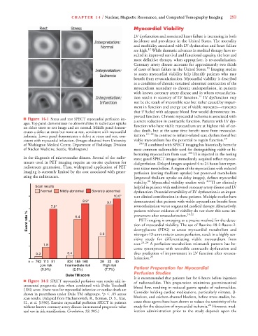
■ Figure 14-1 Stress and rest SPECT myocardial perfusion im- a severe reduction in contractile function. Patients with LV dys-
ages. Top panel demonstrates no abnormalities in radiotracer uptake
on either stress or rest image and are normal. Middle panel demon- function who have viable myocardium are at highest risk of car-
strates a defect at stress but none at rest, consistent with myocardial diac death, but at the same time benefit most from revascular-
15–18
ischemia. Lower panel demonstrates a defect at stress and rest, con- ization. In contrast to infarct-related scar, dysfunctional but
sistent with myocardial infarction. (Images obtained from University viable myocardium has the potential to regain function. 19
of Washington Medical Center, Department of Radiology, Division 201 Tl combined with SPECT imaging has historically been the
of Nuclear Medicine, Seattle, Washington.) most common radionuclide used for distinguishing viable or hi-
bernating myocardium from scar. 201 Tl is injected in the resting
in the diagnosis of microvascular disease. Several of the radio- state; gated SPECT images immediately acquired reflect myocar-
tracers used in PET imaging require an on-site cyclotron for dial perfusion. Delayed images acquired 4 to 24 hours later repre-
radiotracer generation. Thus, widespread application of PET sent tissue metabolism. A region of the myocardium with reduced
imaging is currently limited by the cost associated with gener- perfusion (resting thallium uptake) but preserved metabolism
ating the radiotracers. (improved thallium uptake on delay images), defines myocardial
viability. 20 Myocardial viability studies with 201 Tl are clinically
Scan results helpful in patients with multivessel coronary artery disease and LV
Normal Mildly abnormal Severely abnormal dysfunction. Potential reversibility of LV dysfunction is an impor-
10.0* tant clinical consideration in these patients. Multiple studies have
8.9* demonstrated that patients with viable myocardium benefit from
9.1
7.8* revascularization versus augmented medical therapy. Alternatively,
patients without evidence of viability do not show this same im-
Event rate % 6.4 tion of myocardial viability. The use of fluorine-18-2-fluoro-2-
21,22
provement after revascularization.
PET imaging is emerging as a precise method for the detec-
nitrogen-13-ammonia to assess perfusion, result in a highly sen-
3.6 deoxyglucose (FDG) to assess myocardial metabolism and
sitive study for differentiating viable myocardium from
1.8 scar. 23,24 A perfusion–metabolism mismatch pattern has be-
come synonymous with reversible contractile dysfunction and
0.3 0.4 thus prediction of improvement in LV function after revascu-
larization. 25
n 762 113 51 83 8 68 28 22 40
Low risk Intermediate risk High risk
(0.9%) (2.5%) (7.7%) Patient Preparation for Myocardial
Perfusion Studies
Duke TM score
It is recommended that patients fast for 6 hours before injection
■ Figure 14-2 SPECT myocardial perfusion scan results add in- of radionuclides. This preparation minimizes gastrointestinal
cremental prognostic data when combined with Duke Treadmill blood flow, resulting in reduced gastric uptake of radionuclides.
(TM) score. Event rates for myocardial infarction or cardiac death are
*
shown in parentheses under Duke TM subgroups. *p .05 across Consider holding cardiac medications, particularly nitrates, -
scan results. (Adapted from Hachamovitch, R., Berman, D. S., Kiat, blockers, and calcium-channel blockers, before stress studies, be-
H., et al. [1996]. Exercise myocardial perfusion SPECT in patients cause these agents have been shown to reduce the sensitivity of the
without known coronary artery disease: incremental prognostic value study for the detection of myocardial ischemia. 26 However, med-
and use in risk stratification. Circulation, 93, 905.) ication administration prior to the study depends upon the