
Page 361 - Cardiac Nursing
P. 361
1
1
1
/09
/09
/09
6 A
M
M
2:1
2:1
6 A
/30
87.
q
q
3-3
3-3
87.
6
6
/30
q
xd
xd
Pa
t
ara
ara
p
p
t
In
c.
c.
a
a
In
p
g
g
e 3
Pa
Pa
g
37
A
A
e 3
37
37
33
p
LWB
LWB K34 0-c 16_ p pp333-387.qxd 6/30/09 12:16 AM Page 337 Aptara Inc.
LWBK340-c16_
0-c
16_
33
K34
C HAPTER 1 6 / Arrhythmias and Conduction Disturbances 337
1 1
Table 16-1 ■ CLASSIFICATION OF ANTIARRHYTHMIC DRUGS
Class Action ECG Effect Examples
IA Sodium channel blockade c QRS, c QT Quinidine
2 3 2 C C A 3
Prolong repolarization time Procainamide
Slow conduction velocity Disopyramide
B Suppress automaticity
IB Sodium channel blockade T QT Lidocaine
Accelerate repolarization Mexiletine
IC Sodium channel blockade cc QRS Flecainide
4 4 Marked slowing of Propafenone
A B conduction
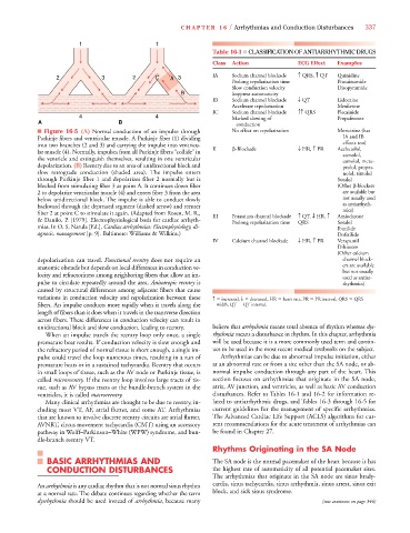
■ Figure 16-5 (A) Normal conduction of an impulse through No effect on repolarization Moricizine (has
Purkinje fibers and ventricular muscle. A Purkinje fiber (1) dividing IA and IB
effects too)
into two branches (2 and 3) and carrying the impulse into ventricu- II -Blockade T HR, c PR Acebutolol,
lar muscle (4). Normally, impulses from all Purkinje fibers “collide” in atenolol,
the ventricle and extinguish themselves, resulting in one ventricular esmolol, meto-
depolarization. (B) Reentry due to an area of unidirectional block and prolol, propra-
slow retrograde conduction (shaded area). The impulse enters nolol, timolol
through Purkinje fiber 1 and depolarizes fiber 2 normally but is Sotalol
blocked from stimulating fiber 3 at point A. It continues down fiber (Other -blockers
2 to depolarize ventricular muscle (4) and enters fiber 3 from the area are available but
below unidirectional block. The impulse is able to conduct slowly not usually used
backward through the depressed segment (dashed arrow) and reenter as antiarrhyth-
mics)
fiber 2 at point C to stimulate it again. (Adapted from Rosen, M. R., III Potassium channel blockade c QT, T HR, c Amiodarone
& Danilo, P. [1979]. Electrophysiological basis for cardiac arrhyth- Prolong repolarization time QRS Sotalol
mias. In O. S. Narula [Ed.], Cardiac arrhythmias: Electrophysiology, di- Ibutilide
agnosis, management [p. 9]. Baltimore: Williams & Wilkins.) Dofetilide
IV Calcium channel blockade T HR, c PR Verapamil
Diltiazem
(Other calcium
depolarization can travel. Functional reentry does not require an channel block-
anatomic obstacle but depends on local differences in conduction ve- ers are available
but not usually
locity and refractoriness among neighboring fibers that allow an im- used as antiar-
pulse to circulate repeatedly around the area. Anisotropic reentry is rhythmics)
caused by structural differences among adjacent fibers that cause
variations in conduction velocity and repolarization between these c increased, T decreased, HR heart rate, PR PR interval, QRS QRS
fibers. An impulse conducts more rapidly when it travels along the width, QT QT interval.
length of fibers than it does when it travels in the transverse direction
across fibers. These differences in conduction velocity can result in
unidirectional block and slow conduction, leading to reentry. believe that arrhythmia means total absence of rhythm whereas dys-
When an impulse travels the reentry loop only once, a single rhythmia means a disturbance in rhythm. In this chapter, arrhythmia
premature beat results. If conduction velocity is slow enough and will be used because it is a more commonly used term and contin-
the refractory period of normal tissue is short enough, a single im- ues to be used in the most recent medical textbooks on the subject.
pulse could travel the loop numerous times, resulting in a run of Arrhythmias can be due to abnormal impulse initiation, either
premature beats or in a sustained tachycardia. Reentry that occurs at an abnormal rate or from a site other than the SA node, or ab-
in small loops of tissue, such as the AV node or Purkinje tissue, is normal impulse conduction through any part of the heart. This
called microreentry. If the reentry loop involves large tracts of tis- section focuses on arrhythmias that originate in the SA node,
sue, such as AV bypass tracts or the bundle-branch system in the atria, AV junction, and ventricles, as well as basic AV conduction
ventricles, it is called macroreentry. disturbances. Refer to Tables 16-1 and 16-2 for information re-
Many clinical arrhythmias are thought to be due to reentry, in- lated to antiarrhythmic drugs, and Tables 16-3 through 16-5 for
cluding most VT, AF, atrial flutter, and some AT. Arrhythmias current guidelines for the management of specific arrhythmias.
that are known to involve discrete reentry circuits are atrial flutter, The Advanced Cardiac Life Support (ACLS) algorithms for cur-
AVNRT, circus movement tachycardia (CMT) using an accessory rent recommendations for the acute treatment of arrhythmias can
pathway in Wolff–Parkinson–White (WPW) syndrome, and bun- be found in Chapter 27.
dle-branch reentry VT.
Rhythms Originating in the SA Node
BASIC ARRHYTHMIAS AND The SA node is the normal pacemaker of the heart because it has
CONDUCTION DISTURBANCES the highest rate of automaticity of all potential pacemaker sites.
The arrhythmias that originate in the SA node are sinus brady-
An arrhythmia is any cardiac rhythm that is not normal sinus rhythm cardia, sinus tachycardia, sinus arrhythmia, sinus arrest, sinus exit
at a normal rate. The debate continues regarding whether the term block, and sick sinus syndrome.
dysrhythmia should be used instead of arrhythmia, because many (text continues on page 346)