
Page 360 - Cardiac Nursing
P. 360
Pa
Pa
M
M
Pa
g
e 3
g
g
6 A
/09
1
/09
/09
1
2:1
6 A
1
2:1
ara
a
t
ara
a
c.
c.
In
In
t
36
36
e 3
36
A
p
p
A
p
33
33
p
3-3
87.
87.
3-3
LWBK340-c16_
LWB
LWB K34 0-c 16_ pp333-387.qxd 6/30/09 12:16 AM Page 336 Aptara Inc.
K34
p
16_
0-c
6
xd
/30
6
/30
q
q
xd
q
336 P A R T III / Assessment of Heart Disease
■ Figure 16-3 Phase 3 block. The ECG on the bot-
tom shows a normal beat followed by a premature
atrial beat that conducts with RBBB. The action po-
tentials on top illustrate that the early beat entered the
right bundle during phase 3, when the membrane po-
tential was still reduced. The resulting action potential
is a slow channel response and conduction fails. (From
Conover, M. [2003]. Understanding electrocardiogra-
phy [8th ed., p. 172]. St. Louis, MO: CV Mosby.)
direction. In addition, conduction velocity must be slow enough rel- a previously depolarized area. Conduction velocity must be slow
ative to tissue refractoriness and circuit length to allow the impulse enough and the refractory period short enough to allow time for
to continue propagating in a circular manner. 3–5 Figure 16-5A the previously stimulated area to recover its ability to conduct. If
illustrates normal conduction of an impulse through an area of the refractory period of the previously stimulated tissue is long or
myocardium, and Figure 16-5B shows reentry occurring as a re- conduction velocity is fast, the impulse dies out because it en-
sult of an area of unidirectional block and slow conduction. 12 counters tissue that is unable to conduct.
For reentry to occur, an area of unidirectional block is neces- Based on these general concepts, three main types of reentry
sary to allow an impulse to conduct in one direction and to pro- have been described. 3–5,13 Anatomic reentry (see Fig. 16-5) in-
vide a return pathway by which the original stimulus can reenter volves an anatomic obstacle around which the circulating wave of
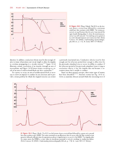
■ Figure 16-4 Phase 4 block. The ECG on the bottom shows a normal beat followed by a pause and a second
beat that conducts with RBBB. The action potential on top illustrates that the pause after the first normal action
potential allowed sufficient time for spontaneous phase 4 depolarization to occur in the RBBB. The impulse after
the pause enters the RBBB at a time when its membrane potential is reduced, resulting in conduction failure.
(From Conover, M. [2003]. Understanding electrocardiography [8th ed., p. 173]. St. Louis, MO: CV Mosby.)