
Page 358 - Cardiac Nursing
P. 358
2:1
2:1
1
1
1
6 A
Pa
Pa
M
6 A
M
/09
xd
xd
q
q
q
6
/09
/09
/30
6
/30
Pa
ara
ara
t
p
t
a
c.
c.
In
a
In
p
e 3
e 3
g
g
g
34
A
p
A
34
34
87.
K34
0-c
16_
LWB K34 0-c 16_ p p pp333-387.qxd 6/30/09 12:16 AM Page 334 Aptara Inc.
LWB
LWBK340-c16_
3-3
3-3
87.
33
33
334 P A R T III / Assessment of Heart Disease
a rhythm due to abnormal automaticity is related to the membrane
potential from which it arose: the less negative the membrane po-
tential (i.e., the greater the depolarization), the faster the rate.
Rhythms due to abnormal automaticity tend to occur at faster
rates than rhythms due to normal automaticity. 1,2
Triggered Activity Due to
Afterdepolarizations
Afterdepolarization is a transient depolarization of the cell mem-
brane that occurs at some time during or right after repolarization
of an action potential. Early afterdepolarizations (EAD) occur
during the repolarization of an action potential. Delayed afterde-
polarizations (DADs) occur after repolarization is complete but
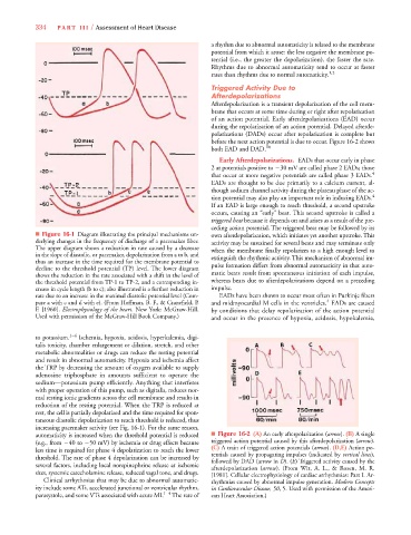
before the next action potential is due to occur. Figure 16-2 shows
both EAD and DAD. 10
Early Afterdepolarizations. EADs that occur early in phase
2 at potentials positive to 30 mV are called phase 2 EADs; those
that occur at more negative potentials are called phase 3 EADs. 4
EADs are thought to be due primarily to a calcium current, al-
though sodium channel activity during the plateau phase of the ac-
tion potential may also play an important role in inducing EADs. 4
If an EAD is large enough to reach threshold, a second upstroke
occurs, causing an “early” beat. This second upstroke is called a
triggered beat because it depends on and arises as a result of the pre-
ceding action potential. The triggered beat may be followed by its
■ Figure 16-1 Diagram illustrating the principal mechanisms un- own afterdepolarization, which initiates yet another upstroke. This
derlying changes in the frequency of discharge of a pacemaker fiber. activity may be sustained for several beats and may terminate only
The upper diagram shows a reduction in rate caused by a decrease when the membrane finally repolarizes to a high enough level to
in the slope of diastolic, or pacemaker, depolarization from a to b, and extinguish the rhythmic activity. This mechanism of abnormal im-
thus an increase in the time required for the membrane potential to pulse formation differs from abnormal automaticity in that auto-
decline to the threshold potential (TP) level. The lower diagram
shows the reduction in the rate associated with a shift in the level of matic beats result from spontaneous initiation of each impulse,
the threshold potential from TP-1 to TP-2, and a corresponding in- whereas beats due to afterdepolarizations depend on a preceding
crease in cycle length (b to c); also illustrated is a further reduction in impulse.
rate due to an increase in the maximal diastolic potential level (Com- EADs have been shown to occur most often in Purkinje fibers
4
pare a with c and d with e). (From Hoffman, B. F., & Cranefield, P . and midmyocardial M cells in the ventricles. EADs are caused
F. [1960]. Electrophysiology of the heart. New York: McGraw-Hill. by conditions that delay repolarization of the action potential
Used with permission of the McGraw-Hill Book Company.) and occur in the presence of hypoxia, acidosis, hypokalemia,
to potassium. 1–4 Ischemia, hypoxia, acidosis, hyperkalemia, digi-
talis toxicity, chamber enlargement or dilation, stretch, and other
metabolic abnormalities or drugs can reduce the resting potential
and result in abnormal automaticity. Hypoxia and ischemia affect
the TRP by decreasing the amount of oxygen available to supply
adenosine triphosphate in amounts sufficient to operate the
sodium—potassium pump efficiently. Anything that interferes
with proper operation of this pump, such as digitalis, reduces nor-
mal resting ionic gradients across the cell membrane and results in
reduction of the resting potential. When the TRP is reduced at
rest, the cell is partially depolarized and the time required for spon-
taneous diastolic depolarization to reach threshold is reduced, thus
increasing pacemaker activity (see Fig. 16-1). For the same reason,
automaticity is increased when the threshold potential is reduced ■ Figure 16-2 (A) An early afterpolarization (arrow). (B) A single
(e.g., from 40 to 50 mV) by ischemia or drug effects because triggered action potential caused by this afterdepolarization (arrow).
less time is required for phase 4 depolarization to reach the lower (C) A train of triggered action potentials (arrow). (D,E) Action po-
s
threshold. The rate of phase 4 depolarization can be increased by tentials caused by propagating impulses (indicated by vertical lines),
followed by DAD (arrow in D). (E) Triggered activity caused by the
E
E
several factors, including local norepinephrine release at ischemic afterdepolarization (arrow). (From Wit, A. L., & Rosen, M. R.
sites, systemic catecholamine release, reduced vagal tone, and drugs. [1981]. Cellular electrophysiology of cardiac arrhythmias: Part I. Ar-
Clinical arrhythmias that may be due to abnormal automatic- rhythmias caused by abnormal impulse generation. Modern Concepts
ity include some ATs, accelerated junctional or ventricular rhythm, in Cardiovascular Disease, 50, 5. Used with permission of the Ameri-
parasystole, and some VTs associated with acute MI. 1–4 The rate of can Heart Association.)