
Page 433 - Cardiac Nursing
P. 433
P
:57
/09
a
P
M
19.
0-4
p40
/30
6
qxd
K34
0-c
18_
LWB K34 0-c 18_ p40 0-4 19. qxd 6 /30 /09 7 7 :57 P M P a g g e e 40 9 A pta ra Inc . .
LWB
LWBK340-c18_p400-419.qxd 6/30/09 7:57 PM Page 409 Aptara Inc.
9 A
40
a
Inc
ra
pta
C HAPTER 1 8 / Cardiac Electrophysiology Procedures 409
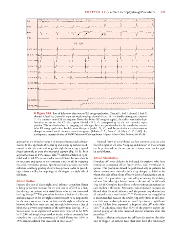
■ Figure 18-4 Loss of delta wave after onset of RF energy application. Channel 1, lead I; channel 2, lead II;
channel 4, lead V 1 ; channel 6, right ventricular tracing; channels 9 and 10, His bundle electrograms; channels
11–15, coronary sinus (CS) electrograms. Notice that before RF energy is applied, the earliest ventricular depo-
larization occurs on the CS electrogram labeled CS 5, 6, corresponding to the left posterior septal
position. This location is where the mapping and ablating catheter was positioned under the mitral valve annulus.
After RF energy application, the delta wave disappears (leads I, II 1 , V 1 ), and the ventricular activation sequence
changes to normal on all coronary sinus electrograms. (Fellows, C. L., Brett, C., & Main, C. C. [1992]. Ra-
diofrequency catheter ablation of Wolff-Parkinson-White syndrome. Virginia Mason Clinic Bulletin, 46, 45–51.)
approach to the mitral annulus is by means of transseptal catheter- Atypical forms of atrial flutter are less common and can arise
ization. In this approach, the ablating and mapping catheter is ad- from the right or left atria. Mapping and ablation of these circuits
vanced to the left atrium through the right heart using a special can be performed but the success rate is lower than that for typi-
sheath assembly to cross the interatrial septum (Fig. 18-5). Both cal atrial flutter.
59
approaches have an 85% success rate. Catheter ablation of right-
sided and septal APs are somewhat more difficult because there is Atrial Fibrillation
no structure analogous to the coronary sinus to aid in mapping Complete AV node ablation is indicated for patients who have
the atrial–ventricular groove. Specialized multielectrode, steerable chronic or paroxysmal AF or flutter with a rapid ventricular re-
catheters, and long guiding sheaths have proven useful in provid- sponse. This procedure should be performed only in patients for
ing catheter stability for mapping and ablating on the right side of whom conventional antiarrhythmic drug therapy has failed or for
the heart. whom the side effects from effective doses of medication are in-
tolerable. This procedure is performed by advancing the ablating
Atrial Flutter catheter from the right femoral vein to the area of the AV node
Primary ablation of classic right atrial isthmus–dependent flutter (Fig. 18-6). Complete heart block with or without a junctional es-
is being performed in most centers and can be offered as a first- cape rhythm is the result. Permanent, rate-responsive pacing is in-
line therapy in patients with atrial flutter who are not interested dicated after AV node ablation, and the patient may discontinue
in drug therapy. 60 This procedure involves ablation of a discrete all antiarrhythmic medications. 49,62 Continuous anticoagulation
anatomic region in the low right atrium thought to be responsible is recommended because the underlying arrhythmia is still pres-
for the macroreentrant circuit. Ablation of the right atrial isthmus ent. Left ventricular dysfunction caused by chronic, rapid heart
between the inferior vena cava and tricuspid valve creates a line of rates in AF has been reported to improve after AV node abla-
block that prevents perpetuation of the arrhythmia (see Fig. 18-7). tion. 63 In addition, more than 80% of the patients report im-
Success rates in an experienced center were reported to be 90% proved quality of life with increased exercise tolerance after this
(n 200). Although the procedure is safe, with an extremely low procedure. 64
complication rate, the recurrence of atrial flutter was 10% to Recent ablation techniques for AF have focused on the abla-
15%. Repeat ablation was successful in most cases. 61 tion of triggers or ectopic beats that arise from the pulmonary