
Page 428 - Cardiac Nursing
P. 428
P
:57
/09
M
a
a
P
19.
0-4
p40
19.
/30
6
qxd
K34
0-c
18_
K34
LWBK340-c18_18_p400-419.qxd 6/30/09 7:57 PM Page 404 Aptara Inc.
LWB
LWB K34 0-c 18_ p40 0-4 19. qxd 6 /30 /09 7 7 :57 P M P a g g e e 40 4 A pta ra Inc . .
40
4 A
pta
pta
Inc
ra
404 P A R T III / Assessment of Heart Disease
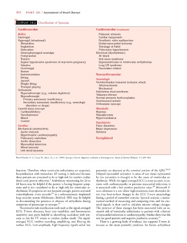
DISPLAY 18-2 Classification of Syncope
Cardiovascular Cardiovascular (continued)
Reflex Pulmonic stenosis
Vasovagal Cardiac tamponade
Vagovagal (situational) Prosthetic valve malfunction
Micturition Global myocardial ischemia
Deglutition Tetralogy of Fallot
Defecation Pulmonary hypertension
Glossopharyngeal neuralgia Electrical (dysrhythmic)
Postprandial AV block
Tussive Sick sinus syndrome
Supine hypotensive syndrome of near-term pregnancy Supraventricular or ventricular arrhythmias
Valsalva Long QT syndrome
Oculovagal Pacemaker related
Sneeze
Instrumentation Noncardiovascular
Diving Neurologic
Jacuzzi Vertebrobasilar transient ischemic attack
Weight lifting Atherosclerosis
Trumpet playing
Mechanical
Orthostatic Subclavian steal syndrome
Hyperadrenergic (e.g., volume depletion) Takayasu disease
Hypoadrenergic Normal pressure hydrocephalus
Primary autonomic insufficiency Unwitnessed seizure
Secondary autonomic insufficiency (e.g., neurologic Orthostatic syncope
disorders or drugs)
Carotid sinus syncope Metabolic
Cardioinhibitory Hypoxia
Vasodepressor Hypoglycemia
Mixed Hyperventilation
Central
Psychiatric
Cardiac Panic disorders
Mechanical (obstructive) Major depression
Aortic stenosis Hysteria
Hypertrophic cardiomyopathy
Pulmonary embolism Unexplained
Aortic dissection
Myocardial infarction
Mitral stenosis
Left atrial myxoma
From Manolis, A. S., Linzer, M., Salem, D., et al. (1990). Syncope: Current diagnostic evaluation and management. Annals of Internal Medicine, 112, 850–863.
function. Therefore, when ventricular arrhythmias are suspected, potentials are detected at the terminal portion of the QRS. 28,29
hospitalization with immediate EP testing is indicated because Delayed myocardial activation in areas of scar tissue represented
these patients are presumed to be at high risk for sudden cardiac by late potentials is thought to be the cause of ventricular ar-
5
death until proven otherwise. Ambulatory monitoring for 24 to rhythmias. While the signal-averaged ECG is most accurate in pa-
48 hours may be helpful if the patient is having frequent symp- tients with cardiomyopathy or previous myocardial infarction, it
toms and is not considered to be at high risk for ventricular ar- is associated with a low positive predictive value. 30 Microvolt T-
rhythmias. If symptoms are not frequent enough, patient-activated wave alternans is a test where high-resolution chest electrodes de-
transtelephonic event recorder 26 or a subcutaneously implanted tect tiny beat-to-beat changes in the ECG T-wave morphology
loop recorder system (Medtronic, Bedford, NH) may be helpful during a period of controlled exercise. Spectral analysis, a mathe-
in documenting the presence or absence of arrhythmia during matical method of measuring and comparing time and the elec-
symptoms of presyncope or syncope. 27 trical signals, is then used to calculate minute voltage changes.
Noninvasive risk stratification tools such as the signal-averaged The presence of these changes has been associated with an in-
ECG, T-wave alternans, heart rate variability, and baroreceptor creased risk of ventricular arrhythmias in patients with a history
sensitivity may prove helpful in identifying candidates with syn- of myocardial infarction or cardiomyopathy. Studies show that the
cope at risk for VT events or sudden cardiac death. The signal- test has good positive and negative predictive accuracy. 31
averaged ECG involves recording, amplifying, and filtering the There is a growing body of evidence that supports T-wave al-
surface ECG. Low-amplitude, high-frequency signals called late ternans as the more powerful predictor for future arrhythmic