
Page 431 - Cardiac Nursing
P. 431
P
:57
/09
M
a
a
P
19.
0-4
p40
19.
/30
6
qxd
K34
0-c
18_
K34
LWB K34 0-c 18_ p40 0-4 19. qxd 6 /30 /09 7 7 :57 P M P a g g e e 40 7 A pta ra Inc . .
L L LWB
LWBK340-c18_18_p400-419.qxd 6/30/09 7:57 PM Page 407 Aptara Inc.
pta
7 A
40
pta
Inc
ra
C HAPTER 1 8 / Cardiac Electrophysiology Procedures 407
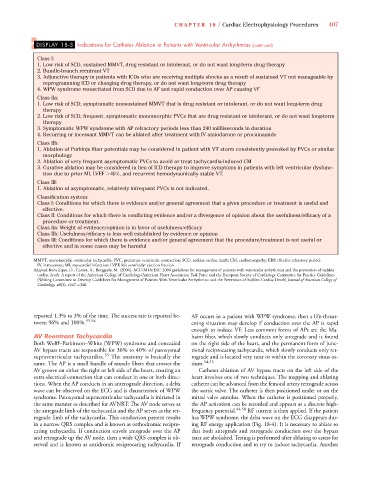
DISPLAY 18-3 Indications for Catheter Ablation in Patients with Ventricular Arrhythmias (continued)
Class I:
1. Low risk of SCD, sustained MMVT, drug resistant or intolerant, or do not want long-term drug therapy
2. Bundle-branch reentrant VT
3. Adjunctive therapy in patients with ICDs who are receiving multiple shocks as a result of sustained VT not manageable by
reprogramming ICD or changing drug therapy, or do not want long-term drug therapy
4. WPW syndrome resuscitated from SCD due to AF and rapid conduction over AP causing VF
Class IIa:
1. Low risk of SCD, symptomatic nonsustained MMVT that is drug resistant or intolerant, or do not want long-term drug
therapy
2. Low risk of SCD, frequent, symptomatic monomorphic PVCs that are drug resistant or intolerant, or do not want long-term
therapy
3. Symptomatic WPW syndrome with AP refractory periods less than 240 millliseconds in duration
4. Recurring or incessant MMVT can be ablated after treatment with IV amiodarone or procainamide
Class IIb:
1. Ablation of Purkinje fiber potentials may be considered in patient with VT storm consistently provoked by PVCs or similar
morphology
2. Ablation of very frequent asymptomatic PVCs to avoid or treat tachycardia-induced CM
3. Curative ablation may be considered in lieu of ICD therapy to improve symptoms in patients with left ventricular dysfunc-
tion due to prior MI, LVEF 40%, and recurrent hemodynamically stable VT.
Class III:
1. Ablation of asymptomatic, relatively infrequent PVCs is not indicated.
Classification system:
Class I: Conditions for which there is evidence and/or general agreement that a given procedure or treatment is useful and
effective.
Class II: Conditions for which there is conflicting evidence and/or a divergence of opinion about the usefulness/efficacy of a
procedure or treatment.
Class IIa: Weight of evidence/opinion is in favor of usefulness/efficacy
Class IIb: Usefulness/efficacy is less well established by evidence or opinion
Class III: Conditions for which there is evidence and/or general agreement that the procedure/treatment is not useful or
effective and in some cases may be harmful
MMVT, monomorphic ventricular tachycardia; PVC, premature ventricular contraction; SCD, sudden cardiac death; CM, cardiomyopathy; ERP, effective refractory period;
IV, intravenous; MI, myocardial infarction; LVEF, left ventricular ejection fraction.
Adapted from Zipes, D., Camm, A., Borggrefe, M. (2006). ACC/AHA/ESC 2006 guidelines for management of patients with ventricular arrhythmias and the prevention of sudden
cardiac death: A report of the American College of Cardiology/American Heart Association Task Force and the European Society of Cardiology Committee for Practice Guidelines
f
(Writing Committee to Develop Guidelines for Management of Patients With Ventricular Arrhythmias and the Prevention of Sudden Cardiac Death) Journal of American College of
Cardiology, 48(5), e247–e346.
reported 1.3% to 3% of the time. The success rate is reported be- AF occurs in a patient with WPW syndrome, then a life-threat-
tween 96% and 100%. 53,54 ening situation may develop if conduction over the AP is rapid
enough to induce VF. Less common forms of APs are the Ma-
AV Reentrant Tachycardia haim fiber, which slowly conducts only antegrade and is found
Both Wolff–Parkinson–White (WPW) syndrome and concealed on the right side of the heart, and the permanent form of junc-
AV bypass tracts are responsible for 30% to 40% of paroxysmal tional reciprocating tachycardia, which slowly conducts only ret-
supraventricular tachycardias. 55 The anatomy is basically the rograde and is located very near or within the coronary sinus os-
same. The AP is a small bundle of muscle fibers that crosses the tium. 54,55
AV groove on either the right or left side of the heart, creating an Catheter ablation of AV bypass tracts on the left side of the
extra electrical connection that can conduct in one or both direc- heart involves one of two techniques. The mapping and ablating
tions. When the AP conducts in an anterograde direction, a delta catheter can be advanced from the femoral artery retrograde across
wave can be observed on the ECG and is characteristic of WPW the aortic valve. The catheter is then positioned under or on the
syndrome. Paroxysmal supraventricular tachycardia is initiated in mitral valve annulus. When the catheter is positioned properly,
the same manner as described for AVNRT. The AV node serves as the AP activation can be recorded and appears as a discrete high-
the antegrade limb of the tachycardia and the AP serves as the ret- frequency potential. 44,58 RF current is then applied. If the patient
rograde limb of the tachycardia. This conduction pattern results has WPW syndrome, the delta wave on the ECG disappears dur-
in a narrow QRS complex and is known as orthodromic recipro- ing RF energy application (Fig. 18-4). It is necessary to ablate so
cating tachycardia. If conduction travels antegrade over the AP that both antegrade and retrograde conduction over the bypass
and retrograde up the AV node, then a wide QRS complex is ob- tract are abolished. Testing is performed after ablating to assess for
served and is known as antidromic reciprocating tachycardia. If retrograde conduction and to try to induce tachycardia. Another