
Page 430 - Cardiac Nursing
P. 430
/30
6
/09
P
:57
0-4
p40
19.
qxd
19.
pta
6 A
pta
Inc
ra
P
M
a
40
a
K34
0-c
18_
K34
LWBK340-c18_18_p400-419.qxd 6/30/09 7:57 PM Page 406 Aptara Inc.
L L LWB
LWB K34 0-c 18_ p40 0-4 19. qxd 6 /30 /09 7 7 :57 P M P a g g e e 40 6 A pta ra Inc . .
406 P A R T III / Assessment of Heart Disease
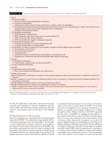
DISPLAY 18-3 Indications for Combined Electrophysiology and Catheter Ablation in Patients with SVT
Class I:
1. Recurrent AVNRT:
a. Poorly tolerated with hemodynamic intolerance
b. Recurrent symptomatic
c. Infrequent or single episode in those who desire complete control of arrhythmia
d. Documented paroxysmal supraventricular tachycardia with only dual AV node pathways or single echo beats on elec-
trophysiological study and no other identified cause of arrhythmia
e. Infrequent well-tolerated AVNRT
2. AP-mediated arrhythmias:
a. WPW syndrome, well tolerated
b. WPW syndrome with rapid conduction or poorly tolerated AF
c. AVRT (concealed AP), poorly tolerated
d. AVRT (concealed AP), single or infrequent episodes
3. Focal atrial tachycardias (AT)
a. Prophylactic therapy for recurrent symptomatic AT
b. Incessant symptomatic or asymptomatic
4. Atrial flutter (if catheter ablation cure not possible, consider AV Node ablation and pacemaker):
a. First episode, well tolerated
b. Recurrent, well tolerated
c. Poorly tolerated
d. Atrial Flutter after IC antiarrhythmic drug (AAD) or amiodarone for AF
e. Symptomatic nonisthmus-dependent atrial flutter after failed AAD therapy
Class IIa:
1. AP-mediated arrhythmias:
a. Preexcitation, asymptomatic (no documented SVT)
2. Focal junctional tachycardia
Class IIb:
1. Inappropriate sinus tachycardia:
a. Sinus node modification/elimination (as a last resort)
Classification system:
Class I: Conditions for which there is evidence and/or general agreement that a given procedure or treatment is useful and
effective.
Class II: Conditions for which there is conflicting evidence and/or a divergence of opinion about the usefulness/efficacy of a
procedure or treatment.
Class IIa: Weight of evidence/opinion is in favor of usefulness/efficacy.
Class IIb: Usefulness/efficacy is less well established by evidence or opinion.
Class III: Conditions for which there is evidence and/or general agreement that the procedure/treatment is not useful or
effective and in some cases may be harmful.
Adapted from Blomstrom-Lundqvist, C., Scheinman, M. M., Aliot, E. M., et al. (2003). ACC/AHA/ESC guidelines for the management of patients with supraventricular
arrhythmias—Executive summary. European Heart Journal, 24(20), 1857–1897; doi: 10.1016.
by APs, AV nodal reentry tachycardia, intra-atrial tachycardias fast pathway, thus perpetuating the reentry circuit and the tachy-
caused by either automatic or reentrant mechanism, atrial fib- cardia. Uncommon or atypical forms of AVNRT can be found
rillation (AF), and atrial flutter. These procedures are also indi- and consist of antegrade fast and retrograde slow pathway con-
cated for some patients with certain types of VT (Display duction, or antegrade and retrograde conduction over multiple
18-3). slow pathway fibers. Ablation of all forms of AVNRT is generally
the same and is accomplished by mapping the slow pathway re-
AV Nodal Reentrant Tachycardia gion, which extends from the posterior/inferior interatrial septum
Dual AV nodal pathways are the substrate for AV nodal reentrant near the coronary sinus ostium up to the anterior/superior intera-
tachycardia (AVNRT). This arrhythmia is responsible for 60% to trial septum. After characteristic electrograms are recorded, RF
44
70% of paroxysmal supraventricular tachycardias. The fast path- energy is applied through the distal ablating electrode (Fig. 18-3).
way has a longer effective refractory period and the slow pathway Repeat programmed stimulation is performed after the ablation in
has a shorter refractory period. The typical form of AVNRT is ini- an attempt to induce the tachycardia. The procedure is considered
tiated when a premature beat from the atrium is blocked in the successful when AVNRT cannot be induced and/or when there is
fast pathway. The early beat conducts down the slow pathway and no evidence of slow pathway conduction. Complete heart block is
then reenters back into the atrium through the fast pathway. This a potentially serious complication because of the close proximity
impulse continues to conduct down the slow pathway and up the of the slow pathway to the compact AV node and has been