
Page 94 - Cardiac Nursing
P. 94
g
P
M
pta
0 A
e 7
009
9/0
9/2
1 A
8:4
0
0-c
03_
L L LWB K34 0-c 03_ p06 9-0 96. qxd 0 9/0 9/2 009 0 0 8:4 1 A M P a a g e 7 0 A pta ra
LWBK340-c03_p069-096.qxd 09/09/2009 08:41 AM Page 70 Aptara
LWB
K34
qxd
ra
96.
p06
9-0
70 PA R T I / Anatomy and Physiology
Higher center
Hypothalamus
Amygdala
Paraventricular cortex
Insular cortex
Anterior cingulate cortex
Mechanoreceptors and
baroreceptors
(Glossopharyngeal) Carotid sinus
Aortic arch
Afferents
Midbrain medula Heart
Nucleus tractus solitarius Lung (cardiopulmonary)
Nucleus ambiguus
Ventrolateral medulla
Parasympathetic
efferents
(vagus)
Spinal cord
Intermediolateral Sympathetic
cell column efferents
Vascular tone
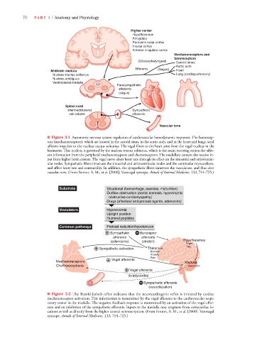
■ Figure 3-1 Autonomic nervous system regulation of cardiovascular hemodynamic responses. The barorecep-
tors (mechanoreceptors), which are located in the carotid sinus, in the aortic arch, and in the heart and lungs, send
afferent impulses to the nucleus tractus solitarius. The vagal fibers to the heart arise from the vagal nucleus in the
brainstem. This nucleus is governed by the nucleus tractus solitarius, which is the main receiving station for affer-
ent information from the peripheral mechanoreceptors and chemoreceptors. The medullary centers also receive in-
put from higher brain centers. The vagal nerve alters heart rate through its effect on the sinoatrial and atrioventric-
ular nodes. Sympathetic fibers innervate the sinoatrial and atrioventricular nodes and the ventricular myocardium,
and affect heart rate and contractility. In addition, the sympathetic fibers innervate the vasculature, and thus alter
vascular tone. (From Fenton, A. M., et al. [2000]. Vasovagal syncope. Annalsof Internal Medicine, 133, 714–725.)
Substrate Situational (hemorrhage, exersise, micturition)
Outflow obstruction (aoritic stenosis, hypertrophic
obstructive cardiomyopathy)
Drugs (afterload and preload agents, adenosine)
Modulators Hypovolemia
Upright position
Humoral peptides
Common pathways Preload reduction/hypotension
Sympathetic Baroceptor
afferents afferents
ypo
ypoth
ypoth
Hyypoth
Hy
ypothalamus
yp
y
po
po
(adenosine) (stretch) Hypot
ight
g
ri
r
F F Fright
F F F F Fear
ear
ear
m
musus
mu
Sympathetic activation Thalammu u s s
Emotiio io o onn n
et
Anxietetty t tyty ty t
Pain
Vagal afferents
Mecchanoreceptors Medulla
a
c
ed
M M M M
e
e
e
m
em
Chemmoreceptoers Vasomo otor
o
Vagal efferents
(bradycardia)
Sympathetic efferents
(vasodilatation)
■ Figure 3-2 The Bezold-Jarisch reflex indicates that the neurocardiogenic reflex is initiated by cardiac
mechanoreceptor activation. This information is transmitted by the vagal afferents to the cardiovascular respi-
ratory center in the medulla. The negative feedback response is transmitted by an activation of the vagal effer-
ents and an inhibition of the sympathetic efferents. Inputs to the medulla may originate from extracardiac lo-
cations as well as directly from the higher central nervous system. (From Fenton, A. M., et al. [2000]. Vasovagal
syncope. Annals of Internal Medicine, 133, 714–725.)