
Page 99 - Cardiac Nursing
P. 99
p06
pta
96.
9-0
5 A
g
ra
e 7
qxd
1 A
8:4
P
M
9/0
0
009
9/2
K34
0-c
03_
L L LWB K34 0-c 03_ p06 9-0 96. qxd 0 9/0 9/2 009 0 0 8:4 1 A M P a a g e 7 5 A pta ra
LWB
LWBK340-c03_p069-096.qxd 09/09/2009 08:41 AM Page 75 Aptara
C HAPTER 3 / Regulation of Cardiac Output and Blood Pressure 75
ble for 10% to 20% of vasodilation in response to hyperther- Adrenergic terminal neuron
mia. 67,68,73 Cholinergic nerves, which innervate the sweat glands, re-
lease a yet to be described co-transmitter that may be functionally
z
Depolarization m 2
a
l
linked to the large and important active cutaneous vasodilation seen adrenerrgic inhibition
Vag l
o
in heat stress. 69,73,74 Additionally, under conditions of hyperthermia, NE
nitric oxide is necessary for the vasodilatory response. 69 Endothelial , A-II
2
nitric oxide (eNOS) is responsible for vasodilation in response to lo- ade NO s i n e
o
adenosine
n
Re-
Re
75
cal cutaneous heating, whereas neuronal nitric oxide (nNOS) is re- uptake NE 2
76
sponsible for vasodilation in response to whole-body heating. The E Angio-II
cutaneous veins constrict in response to local cold and are reflexly 1 A-II Opie (2003)
constricted in response a decrease in skin or core body temperature. 77 2 Vascu ar smooth
la
Nonthermoregulatory control of the cutaneous circulation via m muscle
Vasoconstrict
the arterial and cardiopulmonary baroreflexes plays a role in blood
Vasodilate
pressure control. For example, under normothermic conditions,
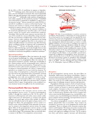
“unloading” of the baroreflex causes cutaneous vasoconstriction. Be- ■ Figure 3-5 Role of neuromodulation in arteriolar constriction
cause of the normally low cutaneous blood flow during normother- and dilation. Norepinephrine is released from the storage granules of
mia, this vasoconstriction contributes little to blood pressure main- the terminal neurons into the synaptic cleft that separates the termi-
tenance. However, under conditions of hyperthermia and during nals from the arterial wall. Norepinephrine has predominantly vaso-
exercise, when there is significant blood flow to the cutaneous vas- constrictive effects acting through postsynaptic 1 receptors. In addi-
tion, norepinephrine stimulates presynaptic 2 receptors to invoke
culature, baroreflex-mediated vasoconstrictive may offset thermoreg- feedback inhibition of its own release, to modulate excess release of
ulatory vasodilation and play an important role in maintenance of NE. Parasympathetic cholinergic stimulation inhibits the release of
blood pressure. 67,73 Of note, the baroreflex sensitivity is not im- norepinephrine and thereby indirectly causes vasodilation. Circulat-
paired by whole-body heating as previously thought; however, heat ing epinephrine stimulates vascular vasodilatory 2 receptors, but also
stress may decrease peripheral vasoconstrictor responsiveness, which presynaptic receptors on the nerve terminal that promotes release of
contributes to an increased susceptibility to orthostatic intolerance. 78 norepinephrine. Angiotensin II, formed ultimately in response to
renin released from the kidneys, is also powerfully vasoconstrictive,
Neurotransmitters acting both by inhibition of norepinephrine release (presynaptic re-
The sympathetic postganglionic fibers that innervate the arterial ceptors, schematically shown to the left of the terminal neuron) and
tree are in general noradrenergic (i.e., release norepinephrine). The also directly on arteriolar receptors. E, epinephrine; NE, norepineph-
rine; A-II, angiotensin II; M2, muscarinic receptor, subtype 2. (From
only exceptions are the postganglionic fibers that innervate the Opie, L. H. [2003]. The heart: Physiology from cell to circulation [4th
sweat glands (sudomotor neurons), which have acetylcholine as ed., p. 25]. Philadelphia: Lippincott Williams & Wilkins.)
their neurotransmitter and the extrapyramidal system, which has
dopamine as the primary neurotransmitter. Norepinephrine is syn-
thesized from tyrosine and is stored in sympathetic nerve terminals.
In response to neuronal stimulation, the “packets” or quanta of nor-
epinephrine are extruded from the axon vesicles by exocytosis. The
vesicular release of norepinephrine is enhanced by angiotensin II Receptors
and cold, whereas the prejunctional effects of potassium, decreased In the parasympathetic nervous system, the nerve fibers are
PO 2 , heat, autacoids (adenosine, bradykinin, serotonin, and cholinergic, which means they liberate acetylcholine. Despite a
prostaglandins), nitric oxide, and acetylcholine inhibit its release 79 common neurotransmitter (acetylcholine), stimulation of various
(Fig. 3-5). The neurotransmitters diffuse over varying small dis- receptors in the parasympathetic nervous system causes different
tances, depending on the width of the junctional cleft, to receptors effects. The reason for the variable response is that there are two
located on effector organs. Norepinephrine is also considered a sys- general types of cholinergic receptors: nicotinic and muscarinic.
temic hormone because of its spillover into the interstitial space. Preganglionic cholinergic receptors, which are found in the sym-
pathetic and parasympathetic nervous systems, are nicotinic. The
Parasympathetic Nervous System nicotinic receptors are located on autonomic ganglia and skeletal
muscle endplates. Stimulation of the nicotinic receptors is excita-
The second branch of the autonomic nervous system is the tory and short-term (milliseconds). These receptors are blocked by
parasympathetic nervous system. The primary parasympathetic curare. In clinical practice, blockade of the nicotinic receptors with
outflow is through four cranial nerves (III, VII, IX, and X). Of various neuromuscular-blocking agents (e.g., succinylcholine, pan-
importance to blood pressure and cardiac output control, cardiac curonium) causes musculoskeletal paralysis (blockade at the skeletal
vagal (cranial nerve X) motorneurons are located in the nucleus muscle endplate) and may potentially cause hypotension because of
ambiguus and dorsal vagal nucleus of the medulla. In addition, blockade at the autonomic ganglia. 80
there are cell bodies located in the spinal cord gray matter at S-2 The primary postganglionic receptor in the heart, smooth
through S-4. Hence, the parasympathetic nervous system is re- muscle, and glandular tissue is muscarinic. These receptors are
ferred to as the craniosacral branch of the autonomic nervous sys- stimulated by muscarine and can be antagonized by atropine and
tem. In contrast to the sympathetic nervous system, the pregan- scopolamine. There are subtypes of the muscarinic receptors that
glionic fibers of the parasympathetic nervous system are long result in varied responses. The primary muscarinic receptors in the
fibers, synapsing on ganglia that are close to or directly attached heart are the muscarinic subtype 2 (M 2 ), which are specifically as-
to the effector organ. The postsynaptic fibers are relatively short, sociated with vagal nerve endings in the heart. The M 2 receptors
in contrast to the fibers of the sympathetic nervous system. have direct and indirect negative inotropic and chronotropic