
Page 96 - Cardiac Nursing
P. 96
P
g
p06
M
pta
ra
e 7
2 A
9-0
8:4
1 A
009
9/2
qxd
96.
9/0
0
K34
0-c
03_
LWB K34 0-c 03_ p06 9-0 96. qxd 0 9/0 9/2 009 0 0 8:4 1 A M P a a g e 7 2 A pta ra
L L LWB
LWBK340-c03_p069-096.qxd 09/09/2009 08:41 AM Page 72 Aptara
72 PA R T I / Anatomy and Physiology
SP fibers nonNMDA receptors SP, GLU neurons
GLU fibers NMDA receptors GLU neurons
GABA fibers NKI receptors GABA neurons Medulla
RVLM
Nodose
ganglion Petrosal
ganglion
NTS
SP
Aortic Carotid
arch sinus
Baroreceptors NA
CVLM
IM
IML
I I I I I I
IM
Parasympathetic Medulla Sympathetic
activity
activity Spinal cord
Heart
Heart
Blood vessels
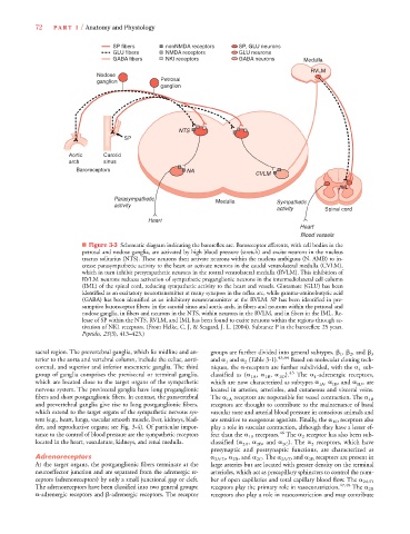
■ Figure 3-3 Schematic diagram indicating the baroreflex arc. Baroreceptor afferents, with cell bodies in the
petrosal and nodose ganglia, are activated by high blood pressure (stretch) and excite neurons in the nucleus
tractus solitarius (NTS). These neurons then activate neurons within the nucleus ambiguus (N. AMB) to in-
crease parasympathetic activity to the heart or activate neurons in the caudal ventrolateral medulla (CVLM),
which in turn inhibit presympathetic neurons in the rostral ventrolateral medulla (RVLM). This inhibition of
RVLM neurons reduces activation of sympathetic preganglionic neurons in the intermediolateral cell column
(IML) of the spinal cord, reducing sympathetic activity to the heart and vessels. Glutamate (GLU) has been
identified as an excitatory neurotransmitter at many synapses in the reflex arc, while gamma-aminobutyric acid
(GABA) has been identified as an inhibitory neurotransmitter at the RVLM. SP has been identified in pre-
sumptive baroreceptor fibers: in the carotid sinus and aortic arch, in fibers and neurons within the petrosal and
nodose ganglia, in fibers and neurons in the NTS, within neurons in the RVLM, and in fibers in the IML. Re-
lease of SP within the NTS, RVLM, and IML has been found to excite neurons within the regions through ac-
tivation of NK1 receptors. (From Helke, C. J, & Seagard, J. L. (2004). Subtance P in the baroreflex: 25 years.
Peptides, 25(3), 413–423.)
sacral region. The prevertebral ganglia, which lie midline and an- groups are further divided into general subtypes, 1 , 2 , and 3
terior to the aorta and vertebral column, include the celiac, aorti- and 1 and 2 (Table 3-1). 43,44 Based on molecular cloning tech-
corenal, and superior and inferior mesenteric ganglia. The third niques, the -receptors are further subdivided, with the 1 sub-
group of ganglia comprises the previsceral or terminal ganglia, classified as ( 1A , 1B , 1D ). 45 The 1 -adrenergic receptors,
which are located close to the target organs of the sympathetic which are now characterized as subtypes 1A , 1B , and 1D , are
nervous system. The previsceral ganglia have long preganglionic located in arteries, arterioles, and cutaneous and visceral veins.
fibers and short postganglionic fibers. In contrast, the paravertebral The 1A receptors are responsible for vessel contraction. The 1B
and prevertebral ganglia give rise to long postganglionic fibers, receptors are thought to contribute to the maintenance of basal
which extend to the target organs of the sympathetic nervous sys- vascular tone and arterial blood pressure in conscious animals and
tem (e.g., heart, lungs, vascular smooth muscle, liver, kidneys, blad- are sensitive to exogenous agonists. Finally, the 1D receptors also
der, and reproductive organs; see Fig. 3-4). Of particular impor- play a role in vascular contraction, although they have a lesser ef-
46
tance to the control of blood pressure are the sympathetic receptors fect than the 1B receptors. The 2 receptor has also been sub-
located in the heart, vasculature, kidneys, and renal medulla. classified ( 2A , 2B , and 2C ). The 2 receptors, which have
presynaptic and postsynaptic functions, are characterized as
Adrenoreceptors 2A/D , 2B , and 2C . The 2A/D and 2B receptors are present in
At the target organs, the postganglionic fibers terminate at the large arteries but are located with greater density on the terminal
neuroeffector junction and are separated from the adrenergic re- arterioles, which act as precapillary sphincters to control the num-
ceptors (adrenoreceptors) by only a small junctional gap or cleft. ber of open capillaries and total capillary blood flow. The 2A/D
The adrenoreceptors have been classified into two general groups: receptors play the primary role in vasoconstriction. 47,48 The 2B
-adrenergic receptors and -adrenergic receptors. The receptor receptors also play a role in vasoconstriction and may contribute