
Page 98 - Cardiac Nursing
P. 98
LWBK340-c03_p069-096.qxd 09/09/2009 08:41 AM Page 74 Aptara
74 PA R T I / Anatomy and Physiology
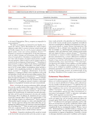
Table 3-1 ■ CARDIOVASCULAR EFFECTS OF AUTONOMIC NERVOUS SYSTEM INNERVATION
Effects
Organ Site Sympathetic Stimulation Parasympathetic Stimulation
Heart Sinoatrial/atrioventricular
Chronotrope ( 1 , 2 ) – Chronotrope
nodes, His-Purkinje system
Myocardium
Inotrope ( 1 , 2 , presynaptic 1 , – Inotrope (minor)
presynaptic 2c )
Coronary arteries Vasoconstriction ( 1D , 2 ) Dilation
Vasodilation ( 2 )
Systemic vasculature Skeletal muscle Vasodilation ( 1 2 , 3 , presynaptic 2 ) —
Vasoconstriction (postsynaptic 2 )
Splanchnic bed Vasoconstriction ( , 2 ) —
Renal Vasoconstriction ( 1 ) —
Cutaneous veins Vasoconstriction (postjunctional 1 , 2 ) Vasodilation
to the onset of hypertension. The 2C receptors are responsible for itance vessels, primarily in the splanchnic bed. The primary trans-
venoconstriction. 46,49 mitter of the vascular smooth muscle sympathetic neuroeffector
The effects of dopamine are mediated by two families of re- junction is norepinephrine. Binding of norepinephrine to the vas-
ceptors (D 1 and D 2 ). The D 1 -like receptors (D 1 and D 5 receptor cular smooth muscle 1 receptor initiates vasoconstriction. The
subtypes) couple with G proteins to activate adenyl cyclase and distribution of the 1 subtypes varies depending on the vascular
the D 2 -like receptors (D 2 , D 3 , and D 4 receptor subtypes) inhibit bed. For example, 1A adrenoreceptors predominate in coronary,
adenyl cyclase release and activate potassium channels. 50,51 splanchnic, renal, and pulmonary vessels, whereas central arteries
Dopamine is a precursor of norepinephrine. In the heart, dopamine and veins express all three 1 receptor subtypes. 46,65 Stimulation
exerts its indirect inotropic and chronotropic effects through the of presynaptic 2 receptors inhibits norepinephrine release and
release of norepinephrine. Stimulation of postjunctional D 1 recep- decreases vasoconstriction, a process called passive vasodilation.
tors in the renal, mesenteric, and splenic arteries produces vasodila- Conversely, stimulation of the postsynaptic 2 receptors, which are
tion and natriuresis. Defects in the D 1 and D 5 receptor may be as- located on large arterioles and perhaps most importantly on the
sociated with the development of hypertension. 50,52 Stimulation of terminal arterioles, causes vasoconstriction. 49 This vasoconstric-
prejunctional D 2 receptors in blood vessels inhibits norepinephrine tion determines the number of open capillaries, and thus capillary
release causing vasodilation. Additionally, in the kidneys stimula- blood flow. The 2 -mediated vasoconstriction of the terminal ar-
tion of the D 2 receptor inhibits norepinephrine release and plays a terioles can be inhibited by metabolic vasodilators (e.g., oxygen,
synergistic role in modulating natriuresis via inhibition of aldos- potassium), particularly in the skeletal muscles. In vascular smooth
terone secretion. 52–55 Exogenous administration of low-dose muscle, the -adrenergic receptors are predominantly of the
dopamine ( 4 g/kg per minute) causes vasodilation of the renal 2 -subtype. Stimulation of these receptors causes vasorelaxation. 44
and splanchnic vascular beds and increases sodium excretion. How-
ever, low-dose dopamine is not reno-protective. 56–58 Intermediate Cutaneous Vasculature
doses of exogenous dopamine (2 to 10 g/kg per minute) stimulate
1 -adrenergic receptors in the heart and increases contractility. Control of the cutaneous circulation arises from both thermoreg-
Higher doses ( 10 g/kg per minute) stimulate -adrenergic ulatory and nonthermoregulatory reflexes. The cutaneous circula-
receptors in the peripheral vasculature and cause vasoconstriction. tion has an extensive distribution of both 1 and 2 adrenorecep-
66
tors, but virtually no adrenoreceptors. The glabrous skin (e.g.,
Heart. In the heart, 1 receptors predominate (80%), although
palms/soles) is innervated only by vasoconstrictive nerves. In con-
there are also a smaller number of 2 receptors (20%), with the 2 trast, nonglabrous skin receives both vasoconstrictive and va-
59
receptors playing a role in coronary vasodilation. Stimulation of sodilator innervations. 67 The sympathetic vasoconstrictor nerves
the 1 and 2 receptors in the heart increases: (1) the rate of dis- release norepinephrine and may also release vasoconstrictive co-
charge of the sinoatrial node, (2) conduction across the atrioventric- transmitters (neuropeptide Y [NPY] or adenosine triphosphate
ular node, and (3) speed of contraction in the atria and ventricles [ATP]), which augments vasoconstriction. 68,69
(chronotropic effect). In addition, 1 stimulation increases cardiac In a thermoneutral environment, the cutaneous resistance vessels
contractility (inotropic effect). There is also a small number ( 1%) in the acral regions (e.g., ears) are tonically constricted, whereas the
of 3 adrenergic receptors in cardiomyocytes. 60 The 3 receptors, nonacral regions (limbs, head, and trunk) have minimal constric-
which mediate negative inotropy via a nitric oxide-dependent path- tion. 70,71 Vasodilation in the acral regions is primarily caused by with-
way, 61 become important during heart failure when they are up- drawal of vasoconstrictive tone (passive vasodilation), whereas vasodi-
regulated and while protective may contribute to functional degrada- lation in nonacral regions is the result of an active process, which is
tion of the failing heart. 60,62 There are small number (approximately sympathetically (but not adrenergically) mediated. Within a “neutral
14%) of 1 receptors located in the atria and ventricles. 63 Stimula- zone,” thermoregulation is controlled entirely by changes in cuta-
tion of the 1 receptors creates a modest inotropic response. 64 72
neous vasomotor tone. An active increase in adrenergic tone causes
Vasculature. Sympathetic stimulation of the arterial tree extends vasoconstriction in response to hypothermia. Conversely, a decrease
to the level of the terminal arterioles and is also present on capac- in adrenergic stimulation causes passive vasodilation and is responsi-