
Page 180 - Color Atlas Of Pathophysiology (S Silbernagl Et Al, Thieme 2000)
P. 180
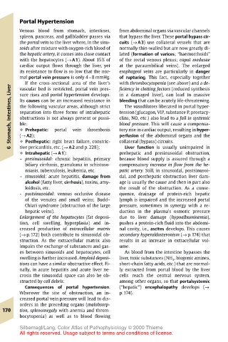
Portal Hypertension
       Venous blood from stomach, intestines,  from abdominal organs via vascular channels
       spleen, pancreas, and gallbladder passes via  that bypass the liver. These portal bypass cir-
       the portal vein to the liver where, in the sinu-  cuits (→ A3) use collateral vessels that are
       soids after mixture with oxygen-rich blood of  normally thin-walled but are now greatly di-
       the hepatic artery, it comes into close contact  lated (formation of varices; “haemorrhoids”
       with the hepatocytes (→ A1). About 15% of  of the rectal venous plexus; caput medusae
       cardiac output flows through the liver, yet  at the paraumbilical veins). The enlarged
       its resistance to flow is so low that the nor-  esophageal veins are particularly in danger
       mal portal vein pressure is only 4–8 mmHg.  of rupturing. This fact, especially together
         If the cross-sectional area of the liver’s  with thrombocytopenia (see above) and a de-
    Liver  vascular bed is restricted, portal vein pres-  ficiency in clotting factors (reduced synthesis
                                       in a damaged liver), can lead to massive
       sure rises and portal hypertension develops.
    Stomach, Intestines,  the following vascular areas, although strict  tension (glucagon, VIP, substance P, prostacy-
       Its causes can be an increased resistance in
                                       bleeding that can be acutely life-threatening.
                                        The vasodilators liberated in portal hyper-
       separation into three forms of intrahepatic
                                       clins, NO, etc.) also lead to a fall in systemic
       obstructions is not always present or possi-
                                       blood pressure. This will cause a compensa-
       ble:
                             thrombosis
                        vein
                                       tory rise in cardiac output, resulting in hyper-
       ! Prehepatic:
                  portal
       ! Posthepatic: right heart failure, constric-
                                       collateral (bypass) circuits.
       tive pericarditis, etc. (→ A2 and p. 228);
                                        Liver function is usually unimpaired in
    6  (→ A2);                         perfusion of the abdominal organs and the
       ! Intrahepatic (→ A1):          prehepatic and presinusoidal obstruction,
       – presinusoidal: chronic hepatitis, primary  because blood supply is assured through a
         biliary cirrhosis, granuloma in schistoso-  compensatory increase in flow from the he-
         miasis, tuberculosis, leukemia, etc.  patic artery. Still, in sinusoidal, postsinusoi-
       – sinusoidal: acute hepatitis, damage from  dal, and posthepatic obstruction liver dam-
         alcohol (fatty liver, cirrhosis), toxins, amy-  age is usually the cause and then in part also
         loidosis, etc.                the result of the obstruction. As a conse-
       - postsinusoidal: venous occlusive disease  quence, drainage of protein-rich hepatic
         of the venules and small veins; Budd–  lymph is impaired and the increased portal
         Chiari syndrome (obstruction of the large  pressure, sometimes in synergy with a re-
         hepatic veins).               duction in the plasma’s osmotic pressure
       Enlargement of the hepatocytes (fat deposi-  due to liver damage (hypoalbuminemia),
       tion, cell swelling, hyperplasia) and in-  pushes a protein-rich fluid into the abdomi-
       creased production of extracellular matrix  nal cavity, i.e., ascites develops. This causes
       (→ p.172) both contribute to sinusoidal ob-  secondary hyperaldosteronism (→ p.174) that
       struction. As the extracellular matrix also  results in an increase in extracellular vol-
       impairs the exchange of substances and gas-  ume.
       es between sinusoids and hepatocytes, cell  As blood from the intestine bypasses the
       swelling is further increased. Amyloid deposi-  liver, toxic substances (NH 3 , biogenic amines,
       tions can have a similar obstructive effect. Fi-  short-chain fatty acids, etc.) that are normal-
       nally, in acute hepatitis and acute liver ne-  ly extracted from portal blood by the liver
       crosis the sinusoidal space can also be ob-  cells reach the central nervous system,
       structed by cell debris.        among other organs, so that portalsystemic
         Consequences of portal hypertension.  (“hepatic”) encephalopathy develops (→
       Wherever the site of obstruction, an in-  p.174).
       creased portal vein pressure will lead to dis-
       orders in the preceding organs (malabsorp-
  170  tion, splenomegaly with anemia and throm-
       bocytopenia) as well as to blood flowing
       Silbernagl/Lang, Color Atlas of Pathophysiology © 2000 Thieme
       All rights reserved. Usage subject to terms and conditions of license.