
Page 196 - Color Atlas Of Pathophysiology (S Silbernagl Et Al, Thieme 2000)
P. 196
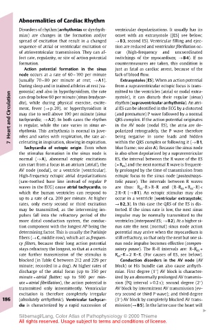
Abnormalities of Cardiac Rhythm
       Disorders of rhythm (arrhythmias or dysrhyth-  ventricular depolarizations. It usually has its
       mias) are changes in the formation and/or  onset with an extrasystole ([ES] see below;
       spread of excitation that result in a changed  → B3, second ES). Ventricular filling and ejec-
       sequence of atrial or ventricular excitation or  tion are reduced and ventricular fibrillation oc-
       of atrioventricular transmission. They can af-  cur  (high-frequency  and  uncoordinated
       fect rate, regularity, or site of action potential  twitchings of the myocardium; → B4). If no
       formation.                      countermeasures are taken, this condition is
         Action potential formation in the sinus  just as fatal as cardiac arrest, because of the
       node occurs at a rate of 60–100 per minute  lack of blood flow.
       (usually 70–80 per minute at rest; → A1).  Extrasystoles (ES). When an action potential
       During sleep and in trained athletes at rest (va-  from a supraventricular ectopic focus is trans-
       gotonia) and also in hypothyroidism, the rate
                                       mitted to the ventricles (atrial or nodal extra-
    Heart and Circulation  dia), while during physical exercise, excite-  QRS complex. If the action potential originates
                                       systole), it can disturb their regular (sinus)
       can drop below 60 per minute (sinus bradycar-
                                       rhythm (supraventricular arrhythmia). An atri-
                                       al ES can be identified in the ECG by a distorted
       ment, fever (→ p. 20), or hyperthyroidism it
                                       (and premature) P wave followed by a normal
       may rise to well above 100 per minute (sinus
       tachycardia; → A2). In both cases the rhythm
                                       in the AV node (nodal ES), the atria are de-
       is regular, while the rate varies in sinus ar-
       niles and varies with respiration, the rate ac-
                                       being negative in some leads and hidden
       celerating in inspiration, slowing in expiration.
                                       within the QRS complex or following it (→ B1,
    7  rhythmia. This arrhythmia is normal in juve-  polarized retrogradely, the P wave therefore
         Tachycardia of ectopic origin. Even when  blue frame; see also A). Because the sinus node
       the stimulus formation in the sinus node is  is also often depolarized by a supraventricular
       normal (→ A), abnormal ectopic excitations  ES, the interval between the R wave of the ES
       can start from a focus in an atrium (atrial), the  (= R ES ) and the next normal R wave is frequent-
       AV node (nodal), or a ventricle (ventricular).  ly prolonged by the time of transmission from
       High-frequency ectopic atrial depolarizations  ectopic focus to the sinus node (postextrasys-
       (saw-toothed base line instead of regular P  tolic pause). The intervals between R waves
       waves in the ECG) cause atrial tachycardia, to  are thus: R ES –R > R–R and (R–R ES + R ES –R) <
       which the human ventricles can respond to  2 R–R (→ B1). An ectopic stimulus may also
       up to a rate of ca. 200 per minute. At higher  occur in a ventricle (ventricular extrasystole;
       rates, only every second or third excitation  → B2,3). In this case the QRS of the ES is dis-
       may be transmitted, as the intervening im-  torted. If the sinus rate is low, the next sinus
       pulses fall into the refractory period of the  impulse may be normally transmitted to the
       more distal conduction system, the conduc-  ventricles (interposed ES; → B2). At a higher si-
       tion component with the longest AP being the  nus rate the next (normal) sinus node action
       determining factor. This is usually the Purkinje  potential may arrive when the myocardium is
       fibers (→ C, middle row), which act as frequen-  still refractory, so that only the next but one si-
       cy filters, because their long action potential  nus node impulse becomes effective (compen-
       stays refractory the longest, so that at a certain  satory pause). The R–R intervals are: R–R ES +
       rate further transmission of the stimulus is  R ES –R = 2 R–R. (For causes of ES, see below).
       blocked (in Table C between 212 and 229 per  Conduction disorders in the AV node (AV
       minute; recorded in a dog). At higher rates of  block) or His bundle can also cause arrhyth-
       discharge of the atrial focus (up to 350 per  mias. First degree (18) AV block is character-
       minute = atrial flutter; up to 500 per min-  ized by an abnormally prolonged AV transmis-
       ute = atrial fibrillation), the action potential is  sion (PQ interval > 0.2 s); second degree (28)
       transmitted only intermittently. Ventricular  AV block by intermittent AV transmission (ev-
       excitation is therefore completely irregular  ery second or third P wave); and third degree
  186  (absolutely arrhythmic). Ventricular tachycar-  (38) AV block by completely blocked AV trans-
       dia is characterized by a rapid succession of  mission (→ B5). In the latter case the heart will
                                                                   "
       Silbernagl/Lang, Color Atlas of Pathophysiology © 2000 Thieme
       All rights reserved. Usage subject to terms and conditions of license.