
Page 193 - Color Atlas Of Pathophysiology (S Silbernagl Et Al, Thieme 2000)
P. 193
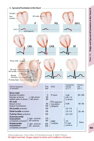
C.  Spread of Excitation in the Heart
                                                                       II
                                                                       Heart
        Excited
        Non               AV node
        excited
                           P               PQ             QRS          Excitation in the
               +
           (mV)  0
               –   ECG (lead II)
                                                                       of
                                                                       Origin and Spread


                      QRS              QRS               ST


                                                                       Plate 7.4
         Sinus node


         AV node                               T
        His bundle
         Bundle
         branches
        Purkinje fibers
                                                   Conduction  Inherent
         Normal sequence            Time  ECG      velocity  rate
         of activation              (ms)           (m·s )   (min )
                                                               –1
                                                      –1
        Sinus node
        Impulse formation            0  P wave     0.05      60–100
        Impulse arrival at  right atrium  50       0.8–1.0
        distant parts of atrium  left atrium  85   in atrium
        AV node                         PQ segment
        Impuls arrival              50  (excitation  0.05    40–55
        Impuls transmission         125  delayed)
        His bundle activated        130            1.0–1.5
        Distal bundle activated     145            1.0–1.5   25–40
        Purkinje fibers activated   150            3.0–3.5
        Subendocardial
        myocardium     right  ventricle  175
        completely activated  left ventricle  190  QRS  1.0
        Subepicardial  right  ventricle  205  complex  in myo-  None
        myocardium     left  ventricle  225        cardium
        completely activated                                          183


       Silbernagl/Lang, Color Atlas of Pathophysiology © 2000 Thieme
       All rights reserved. Usage subject to terms and conditions of license.