
Page 191 - Color Atlas Of Pathophysiology (S Silbernagl Et Al, Thieme 2000)
P. 191
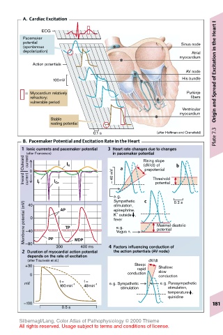
A.  Cardiac Excitation
                                                                       I
                                                                       Heart
               ECG
        Pacemaker
        potential                                           Sinus node
        (spontanous
        depolarization)                                        Atrial
                                                           myocardium  Excitation in the
            Action potentials
                                                             AV node
                  100mV                                     His bundle  of
           Myocardium relatively                             Purkinje
           refractory:                                         fibers
           vulnerable period                                           Origin and Spread
                                                            Ventricular
                                                           myocardium
                 Stable
                 resting potential
                                                                       Plate 7.3
                               0.1 s                (after Hoffman and Cranefield)
        B.  Pacemaker Potential and Excitation Rate in the Heart
        1 Ionic currents and pacemaker potential  3 Heart rate changes due to changes
                                     in pacemaker potential
         Outward (after Francesco)  I K  a     Rising slope  b
          current
                                               (dV/dt) of
                                                prepotential
         Inward  current  0  I f  I Ca  – 40 mV   Threshold
                                                   potential
                                      e.g.      c        0.2 s
                                      Sympathetic
         Membrane potential (mV)  –40 0  AP TP  epinephrine.  Maximal diastolic
          40
                                      stimulation,
                                       +
                                      K  outside   ,
                                      fever
                                       e.g.
                                                    potential
                                       Vagus n.
                 PP
         –80
                   200   MDP 400 ms  4 Factors influencing conduction of
        2 Duration of myocardial action potential  the action potentials (AV node)
          depends on the rate of excitation
          (after Trautwein et al.)               dV/dt
         +30                                Steep:  Shallow:
                                             rapid  slow
           0                              conduction  conduction
         mV         f =  –1  f =  –1  e.g. Sympathetic  e.g. Parasympathetic
                    160min  48min       stimulation         stimulation,
                                                            temperature   ,
                                                            quinidine
         –100                                                         181
                     0.5 s
       Silbernagl/Lang, Color Atlas of Pathophysiology © 2000 Thieme
       All rights reserved. Usage subject to terms and conditions of license.