
Page 205 - Color Atlas Of Pathophysiology (S Silbernagl Et Al, Thieme 2000)
P. 205
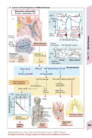
A.  Causes and Consequences of Mitral Stenosis
             Rheumatic endocarditis,
             thrombi, calcification, etc.    ‘P mitrale’
                                        ECG
           1      P LA                   mmHg   P Ao
                                        2
              P Ao                        100
                                                 P LV
                                           50            P LA –P LV
                                                             y
                   P LV                        P LA  a c  v
                                                      x          (after Criley)
                                       Heart  0
        Normal:                        murmur   MOS
        4–6 cm 2                                          MDM PSM
                                                0 0.2 0.4 0.6 0.8 1.0 s
        Mitral                                                         Mitral Stenosis
       opening             Mitral stenosis  Anterior  Interventricular septum
        area                             mitral leaflet
                                           cm
                                           3     E  A      E  F
        Symptoms:           Diastolic .  Echo  D  F  C   D     C
        >2.5 cm 2    : none  flow rate (Q d )  0                       Plate 7.11
              2
        1–2.5 cm  : during              3
                            exercise           Normal  Mitral stenosis
            2
        <1 cm       : at rest  CO        Posterior
                                         mitral leaflet
                                                     Compensation
                 Heart rate  AVD O 2  Left atrial pressure (P LA)
                Stroke volume   LA hypertrophy
                             Left atrial damage  Pulmonary capillary pressure
           Physical exertion,
           fever, pregnancy                  Pulmonary
                         Atrial fibrillation  hypertension
                Heart rate
                                   Atrial   Right heart
                                   thrombi  pressure load
          4 0.7  Heart rate (min )  Brain
                      –1
             60
         Diastolic filling time/time  (min/min)  0.5  (after van der Werf)  Coronaries
                100
                   140


                                   Kidney
           0.3                     Spleen              Pulmonary edema
             Diastolic             Mesentery
             filling time/time     Other
                                   arteries
                                                  Right heart
              Cardiac           Arterial          failure             195
              output            emboli
       Silbernagl/Lang, Color Atlas of Pathophysiology © 2000 Thieme
       All rights reserved. Usage subject to terms and conditions of license.