
Page 206 - Color Atlas Of Pathophysiology (S Silbernagl Et Al, Thieme 2000)
P. 206
Mitral Regurgitation
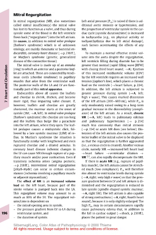
In mitral regurgitation (MR, also sometimes Left atrial pressure (P LA ) is raised if there is ad-
called mitral insufficiency) the mitral valve ditional aortic stenosis or hypertension, and
has lost its function as a valve, and thus during the proportion of ventricular systole in the car-
systole some of the blood in the left ventricle diac cycle (systolic duration/time) is increased
flows back (“regurgitates”) into the left atrium. in tachycardia (e.g., on physical activity or
Its causes, in addition to mitral valve prolapse tachyarrhythmia due to left atrial damage),
(Barlow’s syndrome) which is of unknown such factors accentuating the effects of any
etiology, are mainly rheumatic or bacterial en- MR.
docarditis, coronary heart disease (→ p. 218ff.), To maintain a normal effective stroke vol-
or Marfan’s syndrome (genetic, generalized ume into the aorta despite the regurgitation,
disease of the connective tissue). left ventricle filling during diastole has to be
The mitral valve is made up of an annulus
greater than normal (rapid filling wave [RFW]
Heart and Circulation let are attached. These are connected by tendi- of this increased enddiastolic volume (EDV)
with closing third heart sound; → A). Ejection
(ring) to which an anterior and a posterior leaf-
by the left ventricle requires an increased wall
nous cords (chordae tendineae) to papillary
tension (Laplace’s law), which places a chronic
muscles that arise from the ventricular wall.
load on the ventricle (→ heart failure, p. 224).
The posterior walls of the LA and LV are func-
In addition, the left atrium is subjected to
tionally part of this mitral apparatus.
Endocarditis above all causes the leaflets
high v wave). This causes marked distension
more rigid, thus impairing valve closure. If,
of the left atrium (300–600 mL), while P LA is
7 and chordae to shrink, thicken, and become greater pressure during systole (→ A, left;
however, leaflets and chordae are greatly only moderately raised owing to a long-term
shortened, the murmur starts at the onset of gradual increase in the distensibility (compli-
systole (SM; → A, left). In mitral valve prolapse ance) of the left atrium. As a result, chronic
(Barlow’s syndrome) the chordae are too long MR (→ A, left) leads to pulmonary edemas
and the leaflets thus bulge like a parachute and pulmonary hypertension (→ p. 214)
into the left atrium, where they open. The leaf- much less commonly than mitral stenosis
let prolapse causes a midsystolic click, fol- (→ p.154) or acute MR does (see below). Dis-
lowed by a late systolic murmur (LSM) of re- tension of the left atrium also causes the pos-
flux. In Marfan’s syndrome the situation is terior leaflet of the mitral valve to be displaced
functionally similar with lengthened and even so that the regurgitation is further aggravated
ruptured chordae and a dilated annulus. In (i.e., a vicious circle is created). Another vicious
coronary heart disease ischemic changes in circle, namely MR → increased left heart load
the LV can cause MR through rupture of a pap- → heart failure → ventricular dilation →
illary muscle and/or poor contraction. Even if MR↑↑, can also rapidly decompensate the MR.
transitory ischemia arises (angina pectoris; If there is acute MR (e.g., rupture of papil-
→ p. 218ff.), intermittent mitral regurgitation lary muscle), the left atrium cannot be stretch-
(Jekyll–Hyde) can occur in certain circum- ed much (low compliance). P LA will therefore
stances (ischemia involving a papillary muscle rise almost to ventricular levels during systole
or adjacent myocardium). (→ A, right; very high v wave) so that the pres-
The effect of MR is an increased volume sure gradient between LV and left atrium is di-
load on the left heart, because part of the minished and the regurgitation is reduced in
stroke volume is pumped back into the LA. late systole (spindle-shaped systolic murmur;
This regurgitant volume may amount to as → A, right SM). The left atrium is also capable
much as 80% of the SV. The regurgitant vol- of strong contractions (→ A, right; fourth heart
ume/time is dependent on sound), because it is only slightly enlarged. The
– the mitral opening area in systole, high P LA may in certain circumstances rapidly
– the pressure gradient from LV to LA during cause pulmonary edema that, in addition to
196 ventricular systole, and the fall in cardiac output (→ shock, p. 230ff.),
– the duration of systole. places the patient in great danger.
Silbernagl/Lang, Color Atlas of Pathophysiology © 2000 Thieme
All rights reserved. Usage subject to terms and conditions of license.