
Page 322 - Color Atlas Of Pathophysiology (S Silbernagl Et Al, Thieme 2000)
P. 322
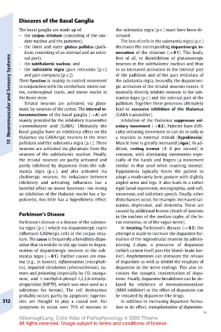
Diseases of the Basal Ganglia
       The basal ganglia are made up of:  the substantia nigra (p.c.) must have been de-
       – the corpus striatum (consisting of the cau-  stroyed.
         date nucleus and the putamen);  The loss of cells in the substantia nigra (p.c.)
       – the inner and outer globus pallidus (palli-  decreases the corresponding dopaminergic in-
         dum, consisting of an internal and an exter-  nervation of the striatum (→ B1). This leads,
    Systems  – the subthalamic nucleus; and  first of all, to disinhibition of glutamatergic
         nal part);
                                       neurons in the subthalamic nucleus and thus
                                       to an increased activation in the internal part
       – the substantia nigra (pars reticulata [p.r.]
         and pars compacta [p.c.]).
                                       of the pallidum and of the pars reticulata of
    Neuromuscular and Sensory  in conjunction with the cerebellum, motor cor-  gic activation of the striatal neurons ceases. It
                                       the substantia nigra. Secondly, the dopaminer-
       Their function is mainly to control movement
       tex, corticospinal tracts, and motor nuclei in
                                       normally directly inhibits neurons in the sub-
                                       stantia nigra (p.r.) and the internal part of the
       the brain stem.
                                       pallidum. Together these processes ultimately
         Striatal neurons are activated, via gluta-
                                       lead to excessive inhibition of the thalamus
       mate, by neurons of the cortex. The internal in-
                                       (GABA transmitter).
       terconnections of the basal ganglia (→ A) are
       mainly provided by the inhibitory transmitter
                                        Inhibition of the thalamus suppresses vol-
                                       culty initiating movement or can do so only as
       basal ganglia have an inhibitory effect on the
       thalamus via GABAergic neurons in the inner
                                       a reaction to external stimuli (hypokinesia).
    10  γ-aminobutyric acid (GABA). Ultimately the  untary movement (→ B2). Patients have diffi-
       pallidum and the substantia nigra (p.r.). These
                                       Muscle tone is greatly increased (rigor). In ad-
       neurons are activated via glutamate from the  dition, resting tremor (4–8 per second) is
       neurons of the subthalamic nucleus. Finally,  common, with alternating movements espe-
       the striatal neurons are partly activated and  cially of the hands and fingers (a movement
       partly inhibited by dopamine from the sub-  similar to that used when counting money).
       stantia nigra (p.c.), and also activated via  Hypokinesia typically forces the patient to
       cholinergic neurons. An imbalance between  adopt a moderately bent posture with slightly
       inhibitory and activating influences has a  angled arms and legs. It also leads to a rather
       harmful effect on motor functions: too strong  rigid facial expression, micrographia, and soft,
       an inhibition of the thalamic nuclei has a hy-  monotone, and indistinct speech. Finally, other
       pokinetic, too little has a hyperkinetic effect.  disturbances occur, for example, increased sal-
                                       ivation, depression, and dementia. These are
                                       caused by additional lesions (death of neurons
       Parkinson’s Disease
                                       in the nucleus of the median raphe, of the lo-
       Parkinson’s disease is a disease of the substan-  cus coeruleus, or of the vagus nerve).
       tia nigra (p.c.) which via dopaminergic tracts  In treating Parkinson’s disease (→ B3) the
       influences GABAergic cells in the corpus stria-  attempt is made to increase the dopamine for-
       tum. The cause is frequently a hereditary dispo-  mation of the nigrostriatal neurons by admin-
       sition that in middle to old age leads to degen-  istering L-dopa, a precursor of dopamine
       eration of dopaminergic neurons in the sub-  (which cannot itself pass the blood–brain bar-
       stantia nigra (→ B1). Further causes are trau-  rier). Amphetamines can stimulate the release
       ma (e.g., in boxers), inflammation (encephali-  of dopamine as well as inhibit the reuptake of
       tis), impaired circulation (atherosclerosis), tu-  dopamine in the nerve endings. This also in-
       mors and poisoning (especially by CO, manga-  creases the synaptic concentration of dopa-
       nese, and 1-methyl-4-phenyl-1,2,3,6-tetrahy-  mine. Finally, dopamine breakdown can be de-
       dropyridine [MPTP], which was once used as a  layed by inhibitors of monoaminooxidase
       substitute for heroin). The cell destruction  (MAO inhibitor) or the effect of dopamine can
       probably occurs partly by apoptosis; superox-  be imitated by dopamine-like drugs.
  312  ides are thought to play a causal role. For  In addition to increasing dopamine forma-
       symptoms to occur, over 70% of neurons in  tion or its effect, transplantation of dopamine-
                                                                   "
       Silbernagl/Lang, Color Atlas of Pathophysiology © 2000 Thieme
       All rights reserved. Usage subject to terms and conditions of license.