
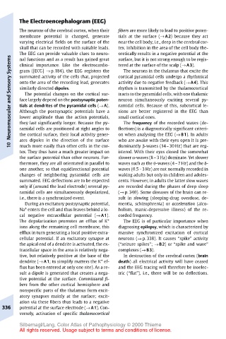
Page 346 - Color Atlas Of Pathophysiology (S Silbernagl Et Al, Thieme 2000)
P. 346
The Electroencephalogram (EEG)
       The neurons of the cerebral cortex, when their  fibers are more likely to lead to positive poten-
       membrane potential is changed, generate  tials at the surface (→ A2) because they act
       varying electrical fields on the surface of the  near the cell body, i.e., deep in the cerebral cor-
       skull that can be recorded with suitable leads.  tex. Inhibition in the area of the cell body the-
       The EEG can provide valuable clues to neuro-  oretically results in a negative potential at the
    Systems  nal functions and as a result has gained great  surface, but it is not strong enough to be regis-
       clinical importance. Like the electrocardio-
                                       tered at the surface of the scalp (→ A3).
                                        The neurons in the thalamus that excite the
       gram ([ECG] → p.184), the EEG registers the
                                       cortical pyramidal cells undergo a rhythmical
       summated activity of the cells that, projected
    Neuromuscular and Sensory  similarly directed dipoles.  rhythm is transmitted by the thalamocortical
                                       activity due to negative feedback (→ A4). This
       onto the area of the recording lead, generates
                                       tracts to the pyramidal cells, with one thalamic
         The potential changes on the cortical sur-
                                       neuron simultaneously exciting several py-
       face largely depend on the postsynaptic poten-
       tials at dendrites of the pyramidal cells (→ A).
                                       ramidal cells. Because of this, subcortical le-
       Although the postsynaptic potentials have a
                                       sions are better registered in the EEG than
                                       small cortical ones.
       lower amplitude than the action potentials,
       they last significantly longer. Because the py-
                                        The frequency of the recorded waves (de-
       the cortical surface, their local activity gener-
                                       on when analysing the EEG (→ B1). In adults
                                       who are awake with their eyes open it is pre-
       ates dipoles in the direction of the surface
    10  ramidal cells are positioned at right angles to  flections) is a diagnostically significant criteri-
                                       dominantly β-waves (14–30 Hz) that are reg-
       much more easily than other cells in the cor-
       tex. They thus have a much greater impact on  istered. With their eyes closed the somewhat
       the surface potential than other neurons. Fur-  slower α-waves (8–3 Hz) dominate. Yet slower
       thermore, they are all orientated in parallel to  waves such as the ϑ-waves (4–7 Hz) and the δ-
       one another, so that equidirectional potential  waves (0.5–3 Hz) are not normally recorded in
       changes of neighboring pyramidal cells are  waking adults but only in children and adoles-
       summated. EEG deflections are to be expected  cents. However, in adults the latter slow waves
       only if (around the lead electrode) several py-  are recorded during the phases of deep sleep
       ramidal cells are simultaneously depolarized,  (→ p. 340). Some diseases of the brain can re-
       i.e., there is a synchronized event.  sult in slowing (sleeping-drug overdose, de-
         During an excitatory postsynaptic potential,  mentia, schizophrenia) or acceleration (alco-
         +
       Na enters the cell and thus leaves behind a lo-  holism, manic-depressive illness) of the re-
       cal negative extracellular potential (→ A1).  corded frequency.
       The depolarization promotes an efflux of K +  The EEG is of particular importance when
       ions along the remaining cell membrane, this  diagnosing epilepsy, which is characterized by
       efflux in turn generating a local positive extra-  massive synchronized excitation of cortical
       cellular potential. If an excitatory synapse at  neurons (→ p. 338). It causes “spike” activity
       the apical end of a dendrite is activated, the ex-  (“seizure spikes”; → B2) or “spike and wave”
       tracellular space in the area is relatively nega-  complexes (→ B3).
       tive, but relatively positive at the base of the  In destruction of the cerebral cortex (brain
                                 +
       dendrite (→ A1; to simplify matters the K ef-  death) all electrical activity will have ceased
       flux has been entered at only one site). As a re-  and the EEG tracing will therefore be isoelec-
       sult a dipole is generated that creates a nega-  tric (“flat”), i.e., there will be no deflections.
       tive potential at the surface. Commissural fi-
       bers from the other cortical hemisphere and
       nonspecific parts of the thalamus form excit-
       atory synapses mainly at the surface; excit-
       ation via these fibers thus leads to a negative
  336  potential at the surface electrode (→ A1). Con-
       versely, activation of specific thalamocortical
       Silbernagl/Lang, Color Atlas of Pathophysiology © 2000 Thieme
       All rights reserved. Usage subject to terms and conditions of license.