
Page 370 - Color Atlas Of Pathophysiology (S Silbernagl Et Al, Thieme 2000)
P. 370
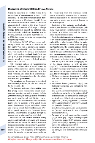
Disorders of Cerebral Blood Flow, Stroke
       Complete cessation of cerebral blood flow  the connection from the dominant hemi-
       causes loss of consciousness within 15–20  sphere to the right motor cortex, is impaired.
       seconds (→ p. 342) and irreversible brain dam-  Bilateral occlusion of the anterior cerebral ar-
       age after seven to 10 minutes (→ A1). Occlu-  tery leads to apathy as a result of damage to
       sion of individual arteries results in deficits in  the limbic system.
    Systems  circumscribed regions of the brain (stroke).  leads to partial contralateral hemianopsia (pri-
                                        Occlusion of the posterior cerebral artery
       The basic mechanism of damage is always
       energy deficiency caused by ischemia (e.g.,
                                       mary visual cortex) and blindness in bilateral
                                       occlusion. In addition, there will be memory
       atherosclerosis, embolism). Bleeding (due to
    Neuromuscular and Sensory  p. 208) also causes ischemia by compressing  cause deficits in the supply area of the anterior
                                       losses (lower temporal lobe).
       trauma, vascular aneurysm, hypertension; →
                                        Occlusion of the carotid or basilar artery can
       neighboring vessels.
                    +
                      +
         By inhibiting Na /K -ATPase, energy defi-
                                       and middle cerebral arteries. When the ante-
       ciency causes the cellular accumulation of
                                       rior choroid artery is occluded, the basal gang-
         +
              2+
                                       lia (hypokinesia), the internal capsule (hemi-
       Na and Ca
                as well as an increased extracel-
                      +
                                       paresis), and optic tract (hemianopsia) are af-
       lular concentration of K , and thus depolariza-
                                       fected. Occlusion of the branches of the poste-
       tion. This results in the cellular accumulation
          –
                                       primarily causes sensory deficits
       also p.10). It also promotes the release of glu-
                                        Complete occlusion of the basilar artery
       tamate, which accelerates cell death via the
              +
                   2+
    10  of Cl , cell swelling, and cell death (→ A; see  rior communicating artery to the thalamus
       entry of Na and Ca .
                                       causes paralysis of all limbs (tetraplegia) and
         Cell swelling, release of vasoconstrictor  of the ocular muscles as well as coma
       mediators, and occlusion of vessel lumina by  (→ p. 342). Occlusion of the branches of the ba-
       granulocytes sometimes prevent reperfusion,  silar artery can cause infarctions in the cerebel-
       despite the fact that the primary cause has  lum, mesencephalon, pons,and medulla oblon-
       been removed. Cell death leads to inflamma-  gata. The effects depend on the site of damage:
       tion that also damages cells at the edge of the  – Dizziness, nystagmus, hemiataxia (cerebel-
       ischemic area (penumbra).        lum and its afferent pathways, vestibular
         The symptoms are determined by the site of  nerve).
       the impaired perfusion, i.e., the area supplied  – Parkinson’s disease (substantia nigra), con-
       by the vessel (→ B).             tralateral hemiplegia and tetraplegia (py-
         The frequent occlusion of the middle cere-  ramidal tract).
       bral artery causes contralateral muscle weak-  – Loss of pain and temperature sensation (hyp-
       ness and spasticity as well as sensory deficits  esthesia or anesthesia) in the ipsilateral half
       (hemianesthesia) by damage to the precentral  of the face and the contralateral limbs (tri-
       and postcentral lateral gyri. Further conse-  geminal nerve [V] and spinothalamic tract).
       quences are ocular deviation (“déviation con-  – Hypacusis (auditory hypesthesia; cochlear
       jugée” due to damage of the visual motor  nerve), ageusis (salivary tract nerve), singul-
       area), hemianopsia (optic radiation), motor  tus (reticular formation).
       and sensory speech disorders (Broca and Wer-  – Ipsilateral ptosis, miosis, and facial anhidro-
       nicke speech areas of the dominant hemi-  sis (Horner’s syndrome, in loss of sympa-
       sphere), abnormalities of spatial perception,  thetic innervation).
       apraxia, and hemineglect (parietal lobe).  – Paralysis of the soft palate and tachycardia
         Occlusion of the anterior cerebral artery  (vagal nerve [X]). Tongue muscle paralysis
       causes contralateral hemiparesis and sensory  (hypoglossal nerve [XII]), drooping mouth
       deficits (due to loss of the medial portion of  (facial nerve [VII]), squinting (oculomotor
       the precentral and postcentral gyri), speech dif-  nerve [III], abducens nerve [VI]).
       ficulties (due to damage of the supplementary  – Pseudobulbar paralysis with global muscu-
  360  motor area) as well as apraxia of the left arm,  lar paralysis (but consciousness maintain-
       when the anterior corpus callosum, and thus  ed).
       Silbernagl/Lang, Color Atlas of Pathophysiology © 2000 Thieme
       All rights reserved. Usage subject to terms and conditions of license.