
Page 48 - Color Atlas Of Pathophysiology (S Silbernagl Et Al, Thieme 2000)
P. 48
Iron Deficiency Anemias
Of the iron (Fe) content in the body (2 g in fe- and plasma), which has a “pocket” for
males, 5 g in males) ca. ⁄3 is bound to hemoglo- 4500 Fe 3+ ions, is a rapidly available iron re-
2
bin (Hb), ⁄4 is stored iron (ferritin, hemosider- serve (ca. 600 mg), while Fe from hemosiderin
1
in), the rest is iron with diverse functions (myo- is more difficult to mobilize (250 mg Fe in
globin, Fe-containing enzymes). Loss of iron is macrophages from liver and bone marrow).
ca. 1 mg/d in males and up to 2 mg/d in fe- Hb-Fe and heme-Fe, released from malformed
males (menstruation, pregnancy, birth). Of Fe erythroblasts (so-called inefficient erythropoi-
taken up in food, 3–15% is absorbed in the esis) and hemolyzed erythroblasts, is bound to
duodenum (→ A); in cases of Fe deficiency it haptoglobin and hemopexin respectively, and
can be up to 25% (see below). Iron intake with taken up by the macrophages in bone marrow
food should therefore be at least 10–20 mg/d or by liver and spleen by endocytosis, 97%
(women > children > men). being reused.
Iron absorption (→ A1). Fe can be absorbed Iron deficiency (serum Fe < 0.4 mg/L; serum
relatively efficiently as heme-Fe 2+ (found in ferritin ↓) inhibits Hb synthesis (→ p. 36) so
meat and fish). The Fe (split off from heme) that hypochromic microcytic anemia devel-
gets into the blood or remains in the mucosa ops: MCH < 26 pg, MCV < 70 fL, Hb < 110 g/L. Its
as ferritin-Fe 3+ and returns to the lumen on causes are (→ A and Table):
mucosal cell disintegration. Non-heme Fe can ! Blood loss (gastrointestinal tract, increased
Blood be absorbed only in the form of Fe , which is menstrual bleeding) in adults is the most com-
2+
+
2+
mon cause of iron deficiency (0.5 mg Fe lost
absorbed by a Fe -H -symport carrier (DCT1)
2+
2+
2+
3 (in competition with Mn , Co , Cd , etc.). A with each mL of blood).
low pH of the chyme is essential for absorption, ! Fe recycling is decreased; this form of ane-
+
because it will 1) increase the H gradient that mia (the second most common worldwide) oc-
drives Fe 2+ into the cell via DCT1, and 2) re- curs with chronic infections. In this situation
lease Fe from compounds in food. Non-heme the Fe regained by the macrophages is no lon-
Fe 3+ in food must be reduced by ferrireductase ger adequately released and thus cannot be re-
(+ascorbate) to Fe 2+ on the surface of the lumi- used.
nal mucosa (→ A1, FR). Fe uptake by blood is ! Fe uptake is too low (malnutrition, especial-
regulated by the intestinal mucosa: in Fe defi- ly in the developing countries).
ciency mucosal ferritin translation is inhibited ! Fe absorption is reduced due to: 1) achlor-
by binding the Fe-regulating protein IRP1 to hydria (atrophic gastritis, after gastrectomy;
ferritin-mRNA, so that more of the absorbed → p.142, 148); and 2) malabsorption in dis-
Fe 2+ can reach the blood. There it is oxidized eases of the upper small intestine or in the
by ceruloplasmin (+copper) to Fe 3+ and bound presence of Fe-binding food components (phy-
to apotransferrin, which transports Fe in plas- tate in cereals and vegetables; tannic acid in
ma (→ A). Transferrin (= apotransferrin with tea, oxalates, etc.).
3+
2 Fe ) is taken up, via transferrin receptors, en- ! There is increased Fe requirement (growth,
docytotically in erythroblasts and in hepatic, pregnancy, breast-feeding).
placental, and other cells. After Fe has trans- ! An apotransferrin defect (rare).
ferred to the target cells, apotransferrin again If Fe overloading occurs in the body, damage
becomes available for Fe absorption from the is caused mainly to the liver, pancreas and
intestine and macrophages (see below). myocardium (hemochromatosis) (→ p. 252).
Iron storage (→ A2). Ferritin (in the intes-
tinal mucosa, liver, bone marrow, erythrocytes,
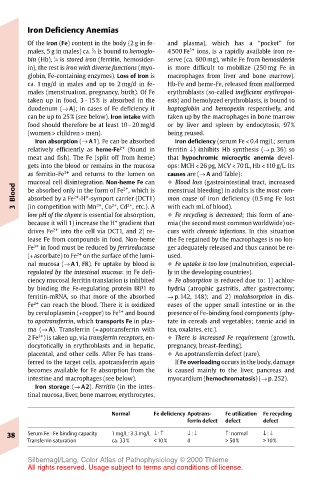
Normal Fe deficiency Apotrans- Fe utilization Fe recycling
ferrin defect defect defect
38 Serum Fe : Fe binding capacity 1 mg/L:3.3 mg/L ↓ : ↑ ↓ : ↓ ↑: normal ↓ : ↓
Transferrin saturation ca. 33% < 10% 0 > 50% > 10%
Silbernagl/Lang, Color Atlas of Pathophysiology © 2000 Thieme
All rights reserved. Usage subject to terms and conditions of license.