
Page 246 - ACCCN's Critical Care Nursing
P. 246
Cardiovascular Alterations and Management 223
of bleeding. Along with vital signs, these are attended patients presenting with chest pain who meet the indica-
every 15 minutes for the first hour, half-hourly for an tions for reperfusion when: (a) facilities are available and
hour, and then hourly according to the patient’s con- can be achieved within 60 minutes; (b) there are contra-
dition, however, patients are advised to report any indications to fibrinolytic therapy described above; (c)
bleeding postdischarge as well. ischaemia would result in large anterior AMI within 4
● ECG monitoring. This includes 12-lead ECG on return hours; or (d) haemodynamic instability or cardiogenic
and ongoing ECG monitoring and chest pain assess- shock are present.
ment to detect reocclusion. Patients need to be
requested to inform nursing staff of any chest pain or A stent is usually inserted to prevent abrupt closure and
26
discomfort. maintain patency for longer. The structure of the stent
● IV anticoagulants such as heparin and/or oral anti- within the vessel enlarges the lumen and prevents vessel
platelet drugs, such as clopidogrel or ticlopidine, may stricture. Restenosis due to intimal hyperplasia is a
be given following thrombolysis to prevent reocclu- relatively common complication, occurring 10–12 weeks
sion in the stent. Assess International Normalised postimplantation. In response to this problem, drug-
Ratio (INR), prothrombin (PT) and partial thrombo- eluting stents have been developed. The drug coatings
plastin time (PTT), as bleeding is more likely to occur include sirolimus, a macrolide antibiotic that has been
if anticoagulants are above the therapeutic range. demonstrated to effectively decrease hyperplasia and
27
prevent reduction of flow. Paclitaxel has also shown
Coronary angioplasty promise in a series of studies. In addition to dactino-
28
Coronary angioplasty (PTCA) procedures are being used mycin, these drugs are undergoing approval processes.
about twice as frequently as coronary artery bypass graft Nursing management of patients post-PTCA includes care
surgery, with 155 PTCA procedures performed for every of the puncture site to prevent bleeding and detect arterial
2
100,000 population in Australia in 2008–09. PTCA rates changes (including clot and aneurysm). The process
29
have grown dramatically in patients aged over 75 years. used to create and maintain access for insertion of the
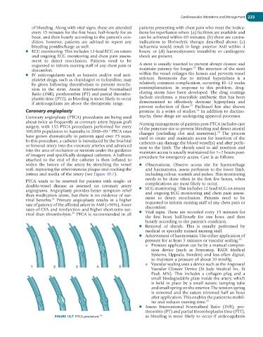
In this procedure, a catheter is introduced by the brachial catheters can damage the blood vessel(s) and alter perfu-
or femoral artery into the coronary arteries and advanced sion to the limb. The sheath used to aid insertion and
into the area of occlusion or stenosis under the guidance maintain access is usually maintained for 1–2 hours post-
of imagery and specifically designed catheters. A balloon procedure for emergency access. Care is as follows:
attached to the end of the catheter is then inflated to
widen the lumen of the artery by stretching the vessel ● Observations. Observe access site for haemorrhage
wall, rupturing the atheromatous plaque and cracking the and haematoma, assess perfusion to the lower limb,
intima and media of the artery (see Figure 10.7). including colour, warmth and pulses. This monitoring
needs to be done often in the first few hours, when
PTCA tends to be reserved for patients with single- or
double-vessel disease as assessed on coronary artery complications are most likely to occur.
angiograms. Angioplasty provides better symptom relief ● ECG monitoring. This includes 12-lead ECG on return
than medication alone, but there is no evidence of sur- and ongoing ECG monitoring and chest pain assess-
vival benefits. Primary angioplasty results in a higher ment to detect reocclusion. Patients need to be
24
rate of patency of the affected artery in AMI (>90%), lower requested to inform nursing staff of any chest pain or
rates of CVA and reinfarction and higher short-term sur- discomfort.
vival than thrombolysis. PTCA is recommended in all ● Vital signs. These are recorded every 15 minutes for
25
the first hour, half-hourly for one hour, and then
hourly according to the patient’s condition.
● Removal of sheath. This is usually performed by
medical or specially trained nursing staff.
● Achievement of haemostasis. Use either application of
pressure for at least 5 minutes or vascular sealing. 29
● Pressure application can be by a manual compres-
sion device (such as Femostop, RADI Medical
Systems, Uppsala, Sweden) and less often digital,
to maintain a pressure of about 20 mmHg.
● Vascular sealing uses a device such as the Angioseal
1 2 3 4 5
Vascular Closure Device (St Jude Medical Inc, St
Paul, MN). This includes a collagen plug and a
small biodegradable plate inside the artery, which
is held in place by a small suture, tamping tube
and small spring on the exterior. The tension spring
is removed and the suture trimmed half an hour
after application. This enables the patient to mobil-
ise and reduces nursing time. 30
● Assess International Normalised Ratio (INR), pro-
thrombin (PT) and partial thromboplastin time (PTT),
FIGURE 10.7 PTCA procedure. as bleeding is more likely to occur if anticoagulants
106