
Page 307 - ACCCN's Critical Care Nursing
P. 307
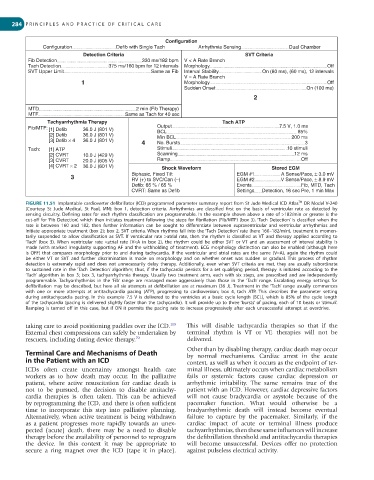
284  P R I N C I P L E S   A N D   P R A C T I C E   O F   C R I T I C A L   C A R E


                                                       Configuration
               Configuration           Defib with Single Tach     Arrhythmia Sensing            Dual Chamber
                            Detection Criteria                                   SVT Criteria
          Fib Detection                        330 ms/182 bpm  V < A Rate Branch
          Tach Detection             375 ms/160 bpm for 12 intervals  Morphology                            Off
          SVT Upper Limit                         Same as Fib  Interval Stability      On (80 ms), (60 ms), 12 intervals
                                                             V = A Rate Branch
                            1                                Morphology                                     Off
                                                             Sudden Onset                            On (100 ms)
                                                                                    2

          MTD                                 2 min (Fib Therapy)
          MTF                             Same as Tach for 40 sec
                 Tachyarrhythmia Therapy                                  Tach ATP
          Fib/MTF:  [1] Defib  36.0 J (801 V)       Output                                  7.5 V, 1.0 ms
                                                                                                  85%
                 [2] Defib  36.0 J (801 V)          BCL                                         200 ms
                                                    Min BCL
                 [3] Defib × 4  36.0 J (801 V)  4   No. Bursts                                       3
          Tach:  [1] ATP                            Stimuli                                    10 stimuli
                 [2] CVRT   10.0 J (429 V)          Scanning                                     12 ms
                 [3] CVRT   20.0 J (605 V)          Ramp                                            Off
                 [4] CVRT × 2  36.0 J (801 V)         Shock Waveform                      Stored EGM
                                                                                             A Sense/Pace, ± 3.0 mV
                        3                   Biphasic, Fixed Tilt              EGM #1         V Sense/Pace, ± 8.9 mV
                                                                              EGM #2
                                            RV (+) to SVC/Can (–)
                                            Defib: 65 % / 65 %                Events               Fib, MTD, Tach
                                            CVRT: Same as Defib               Settings  Detection, 16 sec Pre, 1 min Max
                                                                                                   TM
         FIGURE 11.51  Implantable cardioverter defibrillator (ICD) programmed parameter summary report from St Jude Medical ICD Atlas  DR Model V-240
         (Courtesy  St  Jude  Medical,  St  Paul,  MN):  box  1,  detection  criteria.  Arrhythmias  are  classified  first  on  the  basis  of  ventricular  rate  as  detected  by
         sensing  circuitry.  Defining  rates  for  each  rhythm  classification  are  programmable.  In  the  example  shown  above  a  rate  of  >182/min  or  greater  is  the
         cut-off  for ‘Fib  Detection’,  which  then  initiates  treatment  following  the  steps  for  fibrillation  (Fib/MTF)  (box  3). ‘Tach  Detection’  is  classified  when  the
         rate  is  between  160  and  182,  then  further  information  can  be  sought  to  differentiate  between  supraventricular  and  ventricular  arrhythmias  and
         initiate appropriate treatment (box 2); box 2, SVT criteria. When rhythms fall into the ‘Tach Detection’ rate (here 160–182/min), treatment is momen-
         tarily  suspended  to  allow  classification  as  SVT.  If  ventricular  rate  >atrial  rate,  then  the  rhythm  is  classified  as  VT  and  therapy  applied  according  to
         ‘Tach’  (box  3).  When  ventricular  rate  <atrial  rate  (V<A  in  box  2),  the  rhythm  could  be  either  SVT  or  VT  and  an  assessment  of  interval  stability  is
         made  (with  marked  irregularity  supporting  AF  and  the  withholding  of  treatment).  ECG  morphology  distinction  can  also  be  enabled  (although  here
         is  OFF)  that  compares  morphology  prior  to  and  during  tachycardia.  If  the  ventricular  and  atrial  rates  are  the  same  (V=A),  again  the  rhythm  could
         be  either  VT  or  SVT  and  further  discrimination  is  made  on  morphology  and  on  whether  onset  was  sudden  or  gradual.  This  process  of  rhythm
         detection  is  extremely  rapid  and  does  not  unnecessarily  delay  therapy.  Additionally,  even  when  SVT  criteria  are  met,  they  are  usually  subordinate
         to  sustained  rate  in  the ‘Tach  Detection’  algorithm;  thus,  if  the  tachycardia  persists  for  a  set  qualifying  period,  therapy  is  initiated  according  to  the
         ‘Tach’  algorithm  in  box  3;  box  3,  tachyarrhythmia  therapy.  Usually  two  treatment  arms,  each  with  six  steps,  are  prescribed  and  are  independently
         programmable.  Tachyarrhythmias  in  the  ‘Fib’  range  are  managed  more  aggressively  than  those  in  the  ‘Tach’  range.  Escalating  energy  settings  for
         defibrillation  may  be  described,  but  here  all  six  attempts  at  defibrillation  are  at  maximum  (36  J). Treatment  in  the ‘Tach’  range  usually  commences
         with  one  or  more  attempts  at  antitachycardia  pacing  (ATP),  progressing  to  cardioversion;  box  4,  tach  ATP.  This  describes  the  parameter  setting
         during  antitachycardia  pacing.  In  this  example  7.5  V  is  delivered  to  the  ventricles  at  a  basic  cycle  length  (BCL),  which  is  85%  of  the  cycle  length
         of the tachycardia (pacing is delivered slightly faster than the tachycardia). It will provide up to three ‘bursts’ of pacing, each of 10 beats or ‘stimuli’.
         Ramping  is  turned  off  in  this  case,  but  if  ON  it  permits  the  pacing  rate  to  increase  progressively  after  each  unsuccessful  attempt  at  overdrive.


         taking care to avoid positioning paddles over the ICD.    This  will  disable  tachycardia  therapies  so  that  if  the
                                                         105
         External chest compressions can safely be undertaken by   terminal  rhythm  is  VT  or  VF,  therapies  will  not  be
         rescuers, including during device therapy. 70        delivered.
         Terminal Care and Mechanisms of Death                Other than by disabling therapy, cardiac death may occur
                                                              by  normal  mechanisms.  Cardiac  arrest  in  the  acute
         in the Patient with an ICD                           context, as well as when it occurs as the endpoint of ter-
         ICDs  often  create  uncertainty  amongst  health  care   minal illness, ultimately occurs when cardiac metabolism
         workers  as  to  how  death  may  occur.  In  the  palliative   fails  or  systemic  factors  cause  cardiac  depression  or
         patient,  where  active  resuscitation  for  cardiac  death  is   arrhythmic  irritability.  The  same  remains  true  of  the
         not  to  be  pursued,  the  decision  to  disable  antitachy-  patient with an ICD. However, cardiac depressive factors
         cardia  therapies  is  often  taken.  This  can  be  achieved   will  not  cause  bradycardia  or  asystole  because  of  the
         by reprogramming the ICD, and there is often sufficient   pacemaker  function.  What  would  otherwise  be  a
         time  to  incorporate  this  step  into  palliative  planning.   bradyarrhythmic  death  will  instead  become  eventual
         Alternatively, when active treatment is being withdrawn   failure  to  capture  by  the  pacemaker.  Similarly,  if  the
         as  a  patient  progresses  more  rapidly  towards  an  unex-  cardiac  impact  of  acute  or  terminal  illness  produce
         pected  (acute)  death,  there  may  be  a  need  to  disable   tachyarrhythmias, then these same influences will increase
         therapy before the availability of personnel to reprogram   the defibrillation threshold and antitachycardia therapies
         the  device.  In  this  context  it  may  be  appropriate  to   will  become  unsuccessful.  Devices  offer  no  protection
         secure  a  ring  magnet  over  the  ICD  (tape  it  in  place).   against pulseless electrical activity.