
Page 306 - ACCCN's Critical Care Nursing
P. 306
Cardiac Rhythm Assessment and Management 283
403400 11/11/02 13:20:12 BASELINE 25 mm/sec 0.40
I
II
III
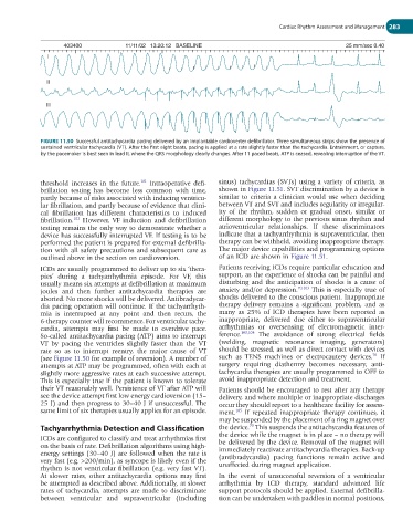
FIGURE 11.50 Successful antitachycardia pacing delivered by an implantable cardioverter defibrillator. Three simultaneous strips show the presence of
sustained ventricular tachycardia (VT). After the first eight beats, pacing is applied at a rate slightly faster than the tachycardia. Entrainment, or capture,
by the pacemaker is best seen in lead II, where the QRS morphology clearly changes. After 11 paced beats, ATP is ceased, revealing interruption of the VT.
threshold increases in the future. Intraoperative defi- sinus) tachycardias (SVTs) using a variety of criteria, as
101
brillation testing has become less common with time, shown in Figure 11.51. SVT discrimination by a device is
partly because of risks associated with inducing ventricu- similar to criteria a clinician would use when deciding
lar fibrillation, and partly because of evidence that clini- between VT and SVT and includes regularity or irregular-
cal fibrillation has different characteristics to induced ity of the rhythm, sudden or gradual onset, similar or
fibrillation. However, VF induction and defibrillation different morphology to the previous sinus rhythm and
102
testing remains the only way to demonstrate whether a atrioventricular relationships. If these discriminators
device has successfully interrupted VF. If testing is to be indicate that a tachyarrhythmia is supraventricular, then
performed the patient is prepared for external defibrilla- therapy can be withheld, avoiding inappropriate therapy.
tion with all safety precautions and subsequent care as The major device capabilities and programming options
outlined above in the section on cardioversion. of an ICD are shown in Figure 11.51.
ICDs are usually programmed to deliver up to six ‘thera- Patients receiving ICDs require particular education and
pies’ during a tachyarrhythmia episode. For VF, this support, as the experience of shocks can be painful and
usually means six attempts at defibrillation at maximum disturbing and the anticipation of shocks is a cause of
joules and then further antitachycardia therapies are anxiety and/or depression. 70,103 This is especially true of
aborted. No more shocks will be delivered. Antibradycar- shocks delivered to the conscious patient. Inappropriate
dia pacing operation will continue. If the tachyarrhyth- therapy delivery remains a significant problem, and as
mia is interrupted at any point and then recurs, the many as 25% of ICD therapies have been reported as
6-therapy counter will recommence. For ventricular tachy- inappropriate, delivered due either to supraventricular
cardia, attempts may first be made to overdrive pace. arrhythmias or oversensing of electromagnetic inter-
So-called antitachycardia pacing (ATP) aims to interrupt ference. 103,104 The avoidance of strong electrical fields
VT by pacing the ventricles slightly faster than the VT (welding, magnetic resonance imaging, generators)
rate so as to interrupt reentry, the major cause of VT should be stressed, as well as direct contact with devices
70
(see Figure 11.50 for example of reversion). A number of such as TENS machines or electrocautery devices. If
attempts at ATP may be programmed, often with each at surgery requiring diathermy becomes necessary, anti-
slightly more aggressive rates at each successive attempt. tachycardia therapies are usually programmed to OFF to
This is especially true if the patient is known to tolerate avoid inappropriate detection and treatment.
their VT reasonably well. Persistence of VT after ATP will Patients should be encouraged to rest after any therapy
see the device attempt first low energy cardioversion (15– delivery, and where multiple or inappropriate discharges
25 J) and then progress to 30–40 J if unsuccessful. The occur they should report to a healthcare facility for assess-
same limit of six therapies usually applies for an episode. ment. If repeated inappropriate therapy continues, it
105
may be suspended by the placement of a ring magnet over
70
Tachyarrhythmia Detection and Classification the device. This suspends the antitachycardia features of
ICDs are configured to classify and treat arrhythmias first the device while the magnet is in place – no therapy will
be delivered by the device. Removal of the magnet will
on the basis of rate. Defibrillation algorithms using high- immediately reactivate antitachycardia therapies. Back-up
energy settings (30–40 J) are followed when the rate is (antibradycardia) pacing functions remain active and
very fast (e.g. >200/min), as syncope is likely even if the unaffected during magnet application.
rhythm is not ventricular fibrillation (e.g. very fast VT).
At slower rates, other antitachycardia options may first In the event of unsuccessful reversion of a ventricular
be attempted as described above. Additionally, at slower arrhythmia by ICD therapy, standard advanced life
rates of tachycardia, attempts are made to discriminate support protocols should be applied. External defibrilla-
between ventricular and supraventricular (including tion can be undertaken with paddles in normal positions,