
Page 544 - ACCCN's Critical Care Nursing
P. 544
Gastrointestinal, Liver and Nutritional Alterations 521
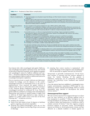
TABLE 19.11 Treatment of liver failure complications
Condition Treatment
Hepatic encephalopathy ● Treatment revolves around general supportive therapy until liver function recovers or liver transplant is
undertaken. 236,256
● Cerebral oedema and raised intracranial pressure are treated as for an acute head injury (see Ch 17).
● Reduce production and absorption of ammonia by preventing/controlling upper gastrointestinal bleeding and
gastrointestinal administration of non-adsorbable disaccharides such as lactulose or lactitol to remove protein
derived from dietary intake or bleeding. 274
Hepatorenal syndrome ● Liver transplant is the primary treatment for type 1 HRS in patients with cirrhosis.
(HRS) ● If transplant is contraindicated or delayed, vasocontrictors (e.g. terlipressin) may be effective in constricting the
dilated splanchnic arterial bed, thus improving renal perfusion pressure and renal function. Vasocontrictors may
be given in association with intravenous albumin in order to increase intravascular volume. 262,263
Variceal bleeding A successful outcome, as in all cases of gastrointestinal haemorrhage, hinges on prompt resuscitation,
haemodynamic support, and correction of haemostatic dysfunction, preferably in the intensive care setting.
● The patient is intubated for airway protection.
● Adequate IV access in inserted, preferably large, wide-bore cannulas for rapid fluid resuscitation.
● Haemodynamic instability is corrected with volume expanders initially and then blood products.
• The source of bleeding is identified by endoscope, and varices are banded/ligated (latex bands placed around the
varices to ‘strangle’ the vessel), or sclerotherapy or diathermy (heat used to cauterise bleeding vessel) is used.
● Terlipressin and octreotide infusions may be used to reduce portal circulation pressure.
● If bleeding is uncontrollable, a balloon tamponade device is inserted.
Ascites Salt and water restrictions along with diuretic therapy are methods that have been used to control ascites in the
preliminary phases of end-stage liver failure; however, in the intensive care setting these measures are impractical
and usually unsuccessful.
● Paracentesis is very effective at reducing ascites and is a simple procedure to remove fluid and an aid in diagnosis.
● Correction of coagulopathy or thrombocytopenia should be considered when the INR is greater than 2.5 or the
platelet count markedly reduced.
● Paracentesis may aid in determining the cause of ascites (ascites-serum albumin gradient, ascitic cytology,
microscopy and culture for acid-fast bacilli, chylous ascites) and in establishing or excluding primary or secondary
peritonitis in patients with ascites (ascitic WCC and neutrophil count, culture).
● Litres of ascites are normally removed, and the volume is replaced with IV concentrated albumin to prevent fluid
shifts and hypotension.
● Mean arterial pressures, central venous pressures, heart rate and urine output are carefully monitored during the
procedure. For every litre of ascites removed, 6–8 g albumin is infused. 275
four-lumen tube with oesophageal and gastric balloons, ● ensuring that correct traction is maintained, with
and oesophageal and gastric aspiration ports. The benefit regular checking of tube migration and checking posi-
of this tube is that direct pressure can be applied on gastric tion at nares/lips at regular intervals (4/24 hours).
and oesophageal varices by balloon inflation and trac- Tamponade is generally maintained for 24–48 hours,
tion. 276 The Linton tube has one lumen for inflation of the then traction is removed and the balloon deflated to
pear-shaped gastric balloon and two additional lumens for assess for further bleeding. If the patient is
oesophageal and gastric aspiration.
stabilised, endoscopy can be performed. If bleeding
Prior to insertion (oral or nasal), balloons are lubricated, persists, the balloon(s) is/are reinflated and traction
checked for leakage, and the distance to the cardio- reapplied. 264,276
oesophageal junction is estimated (nose to ear, then to
xiphisternum). Once inserted, the gastric balloon is Once the patient has been stabilised, a transjugular intra-
inflated with 50 mL air and pulled back until resistance hepatic portosystemic stent/shunt (TIPS) may be con-
is felt. Position (lying compressed against the cardio- sidered to control variceal haemorrhage. TIPS is a metal
oesophageal junction) is confirmed by X-ray. Then the expandable stent inserted to decompress the portal
277
gastric balloon is inflated according to the manufacturer’s venous system.
instructions and traction is applied using a weight (500 Extracorporeal liver support
or 1000 mL IV fluid bag) attached to rope; traction is
applied via a pulley and IV pole at the foot of the bed. The aim of extracorporeal liver support therapy is to allow
Nursing care 276 of patients involves: time for liver recovery or to provide support until a
234
liver transplant is possible. Either biological or non-
● sedation for comfort biological systems are available for liver support. Biologi-
● head of the bed raised at least 30 degrees to facilitate cal systems utilise pig hepatocytes or hepatoma cells to
gastric emptying and prevent aspiration achieve removal of toxins, 234 but this requires complex
● ensuring that gastric/oesophageal ports are on free technical support in specialist centres. Non-biological
drainage, with regular monitoring of type and amount systems are similar to renal replacement circuits, and use
of drainage albumin as a dialysis medium or dialyse against an