
Page 564 - ACCCN's Critical Care Nursing
P. 564
Management of Shock 541
shock. Therapy is targeted to maintain oxygen delivery
TABLE 20.2 Lactate production 9 (DO 2 ) to vital organs to prevent ischaemia and cell
death. 25,26 Ideally, organ systems and tissues should be
25
Lactate production monitored individually, however global measures such
as perfusion pressure, cardiac output (CO) and DO 2 ,
Product of carbohydrate Glucose, glycolysis; pyruvate,
metabolism (1400– lactate are commonly used as surrogates to assist in treatment
19
4500 mmol/day) decision making. Patient assessment and haemody-
namic monitoring, including calculation of CO, are
Rise in lactate levels
used to differentiate shock states and assess progress
Tissue hypoxia Hypodynamic shock in relation to treatment. 26–28 CO is seen by many clini-
Organ ischaemia cians as an important assessment of shocked patients
Hypermetabolism Increased aerobic glycolysis as it is a major determinant of DO 2 . 25,26 Critically ill
Increased protein catabolism patients are frequently assessed clinically, although
Haematological malignancies cardiac output estimations from physical examination
Decreased clearance of lactate Liver failure are generally unreliable and patient status may change
29
Shock quickly. Therefore invasive techniques are most com-
monly used in critical care to measure CO (see also
Inhibition of pyruvate Thiamine deficiency
dehydrogenase Endotoxin Chapter 9).
Activation of inflammatory cells
Phagocytosis Wounds (e.g. trauma/burns) NON-INVASIVE ASSESSMENT
Liver
Gastrointestinal Perfusion status is determined clinically using gross organ
Lungs (e.g. ARDS) function such as mental status, urine output and periph-
6
Major source in sepsis eral warmth and colour. Basic physical assessment of
cardiovascular, central nervous system and renal function
Phagocytes Lungs are essential when assessing a patient at risk of shock.
Wounds Subtle changes in urine output, heart rate and capillary
Liver: neutrophil sequestration
increased, glucose uptake refill are all signs of physiological compensation in
increased response to altered tissue perfusion associated with shock.
Gut: prone to hypoxia, Regular tracking of these vital signs and trend monitoring
phagocytes through careful documentation can alert clinicians to
impending deterioration in the shock state. Level of con-
sciousness may deteriorate; an early sign may be anxiety,
and specific management principles to avoid, or at least and progress to restlessness, agitation or coma. Other
limit, tissue injury and the eventual progression to organ assessment findings include cool, clammy skin, postural
3
failure. hypotension, tachycardia and decreased urine output.
The reliability of these measures is questionable, particu-
PATIENT ASSESSMENT larly where multiple assessments by different clinicians
are performed; in the ICU continuous ECG monitoring
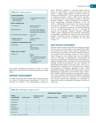
Critically ill patients often exhibit signs of tissue hypoxia and invasive monitoring techniques are employed to
25
as a result of cardiovascular disturbances. Table 20.3 assist in the objective assessment of changes in cardiovas-
provides an overview of the physiological changes in cular state.
TABLE 20.3 Physiological changes in shock 37
Physiological change
Shock Systemic vascular Pulmonary capillary Pulmonary vascular
classification Cardiac output resistance Capillary circulation pressure resistance
Hypovolaemic ↓ ↑ ↓ ↓ ↑
Cardiogenic ↓ ↑ ↓ ↑ ↑
Distributive:
● septic ↑ ↓ ↓ ↓ ↑
● anaphylactic ↓ ↓ ↓ ↓ ↓
● neurogenic ↓/= ↓ ↓ ↓ ↑
Obstructive ↓ ↓ ↓ ↑ ↑
↑ increase; ↓ decrease; = no change.