
Page 565 - ACCCN's Critical Care Nursing
P. 565
542 P R I N C I P L E S A N D P R A C T I C E O F C R I T I C A L C A R E
Although CO estimations based on physical assessment
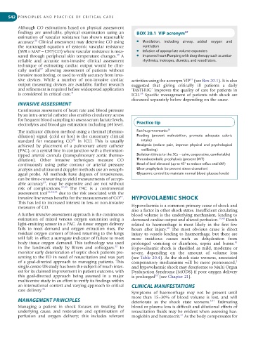
findings are unreliable, physical examination using an BOX 20.1 VIP acronym 37
estimation of vascular resistance has shown reasonable
30
accuracy. Clinical assessment may determine CO using ● Ventilation, including airway, added oxygen and
the rearranged equation of systemic vascular resistance ventilation
(SVR = MAP − CVP/CO) where vascular resistance is mea- ● Infusion of appropriate volume expanders
30
sured through peripheral skin temperature changes. A ● Improved heart Pumping with drug therapy such as antiar-
reliable and accurate non-invasive clinical assessment rhythmics, inotropes, diuretics, and vasodilators.
technique of estimating cardiac output would be clini-
27
cally useful allowing assessment of patients without
invasive monitoring, or used to verify accuracy from inva-
sive devices. While a number of non-invasive cardiac activities using the acronym VIP (see Box 20.1). It is also
37
output measuring devices are available, further research suggested that giving critically ill patients a daily
and refinement is required before widespread application ‘FASTHUG’ improves the quality of care for patients in
is considered in critical care. 31 ICU. Specific management of patients with shock are
38
discussed separately below depending on the cause.
INVASIVE ASSESSMENT
Continuous assessment of heart rate and blood pressure
by an intra-arterial catheter also enables circulatory access
for frequent blood sampling to assess serum lactate levels,
electrolytes and blood gas estimation including pH level. Practice tip
The indicator dilution method using a thermal (thermo- Fast hug mnemonic: 38
dilution) signal (cold or hot) is the customary clinical Feeding (prevent malnutrition, promote adequate caloric
26
standard for measuring CO in ICU. This is usually intake)
achieved by placement of a pulmonary artery catheter Analgesia (reduce pain, improve physical and psychological
(PAC), or a central line in conjunction with a thermistor- wellbeing)
tipped arterial cannula (transpulmonary aortic thermo- Sedation (titrate to the 3Cs – calm, cooperative, comfortable)
dilution). Other invasive techniques measure CO Thromboembolic prophylaxis (prevent DVT)
continuously using pulse contour or arterial pressure Head of bed elevated (up to 45° to reduce reflux and VAP)
analysis and ultrasound doppler methods use an oesoph- Ulcer prophylaxis (to prevent stress ulceration)
ageal probe. All methods have degrees of invasiveness, Glycaemic control (to maintain normal blood glucose levels)
can be time-consuming to yield measurements of accept-
able accuracy , may be expensive and are not without
32
risk of complications. 27,33 The PAC is a controversial
assessment tool 26,28,33 due to the risk associated with the
invasive line versus benefits for the measurement of CO . HYPOVOLAEMIC SHOCK
34
This has led to increased interest in less or non-invasive
measures of CO. Hypovolaemia is a common primary cause of shock and
also a factor in other shock states. Insufficient circulating
A further invasive assessment approach is the continuous blood volume is the underlying mechanism, leading to
estimation of mixed venous oxygen saturation using a decreased cardiac output and altered perfusion. 39,40 Death
light-emitting sensor in a PAC. As tissue oxygen delivery related to haemorrhage is most likely in the first few
fails to meet demand and oxygen extraction rises, the hours after injury. The most obvious cause is direct
40
residual oxygen content of blood returning to the lungs injury to vessels leading to haemorrhage, but there are
will fall; in effect a surrogate indicator of failure to meet more insidious causes such as dehydration from
body tissue oxygen demand. This technology was used prolonged vomiting or diarrhoea, sepsis and burns.
41
35
in the landmark study by Rivers and colleagues. to Hypovolaemic shock is classified as mild, moderate or
monitor early deterioration of septic shock patients pre- severe, depending on the amount of volume loss
senting to the ED in need of resuscitation and was part (see Table 20.4). As the shock state worsens, associated
3
of a goal-directed approach to managing patients. This compensatory mechanisms will be more pronounced,
single-centre US study has been the subject of much inter- and hypovolaemic shock may deteriorate to Multi Organ
est for its claimed improvement in patient outcome, with Dysfunction Syndrome (MODS) if poor oxygen delivery
this goal-directed approach being assessed in a major is prolonged (see Chapter 21).
39
multicentre study in an effort to verify its findings within
an international context and varying approach to critical CLINICAL MANIFESTATIONS
care delivery. 36
Symptoms of haemorrhage may not be present until
more than 15–30% of blood volume is lost, and will
MANAGEMENT PRINCIPLES deteriorate as the shock state worsens. 3,41 Estimating
Managing a patient in shock focuses on treating the blood or plasma loss is difficult and dilutional effects of
underlying cause, and restoration and optimisation of resuscitation fluids may be evident when assessing hae-
41
perfusion and oxygen delivery; this includes relevant moglobin and hematocrit. As the body compensates for