
Page 586 - ACCCN's Critical Care Nursing
P. 586
Multiple Organ Dysfunction Syndrome 563
PATHOPHYSIOLOGY breakdown of cellular components into apoptic bodies.
This normally orderly process is deranged in critical
The syndrome of multiple organ dysfunction is most illness, leading to tissue or organ bed injury and MODS.
closely related to an outcome of sepsis, which was Proinflammatory cytokines released in sepsis may delay
described in Chapter 20. MODS is a state characterised apoptosis in activated macrophages and neutrophils, but
by aberrant cellular responses involving multiple organ in other tissues, such as gut endothelium, accelerated
systems and sequential processes. The pathogenesis of apoptosis occurs. 8
MODS is complex, simultaneously involving every cell
type, neuro-hormonal axis and organ system. 7 In contrast, necrosis is a form of cell death characterised
by cellular swelling and loss of membrane integrity as a
In brief, hypoxic hypoxia results from altered metabolic result of hypoxia or trauma. Necrosis has been termed
regulation of tissue oxygen delivery which contributes to ‘cellular energy crisis’, and is unregulated resulting in
10
further organ dysfunction. Microcirculatory injury as a loss of membrane sodium/potassium/ATP-ase pumps.
result of lytic enzymes, and vasoactive substances (nitric This loss leads to cell swelling, rupture and spillage of
oxide, endothelial growth factor), is compounded by the intracellular contents into surrounding regions creating
inability of erythrocytes to navigate the septic microcir- collateral damage. Necrosis therefore can involve signifi-
10
culation. Mitochondrial electron transport is affected by cant amounts of tissue and organ bed damage. Apoptosis
endotoxins in sepsis, nitric oxide and TNF-alpha, leading differs from necrosis in that it does not seem to involve
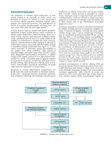
to disordered energy metabolism (see Figure 21.1). This the recruitment of inflammatory cells or mediators to
causes cytopathic or histotoxic anoxia (the inability to complete its task. Activation of an enzyme cascade sys-
8
use oxygen, even when available). This context of tematically cleaves proteins, including the cell’s nuclear
7,8
impaired oxygen utilisation rather than delivery results DNA, with the end-result being death of the cell. This
from diminished mitochondrial production of cellular requires energy from mitrochondria and if not available
energy (ATP), despite normal or even supranormal intra- necrosis of the cell occurs. Apoptosis and necrosis are
9
cellular PO 2 levels. Cytopathic hypoxia appears resistant processes that if is therefore important to understand in
to resuscitation measures, and this may ultimately worsen relation to future MODS research.
already-existing organ dysfunction. During sepsis or isch-
aemia, mitochondria respond by facilitating cell death Increased concentrations of cell-free plasma DNA are
rather than the restoration of homeostasis. 7 present in various clinical conditions such as stroke, myo-
cardial infarction and trauma, a likely result of acceler-
Apoptosis is normal physiological programmed cell ated cell death. Maximum plasma DNA concentrations
death and is the main mechanism to eliminate dysfunc- correlated significantly with APACHE II scores and
10
tional cells. Apoptosis involves chromatin condensa- maximum SOFA scores (described later in this chapter),
tion, membrane blebbing, cell shrinkage and subsequent with cell-free plasma DNA concentrations higher in
Reduced delivery of
oxygen and glucose
Anaerobic metabolism Protein synthesis
Lactate ATP production Lipolysis
pH Cell function
+
+
Na /K pump
+
Intracellular Ca ++ K + Na and H O
2
Denaturing of protein
Cell membrane leakiness Cellular swelling
Lysis and rupture
of organelles
Cell lysis/death
Protein synthesis
Lipolysis
Cell function
FIGURE 21.1 Pathophysiology of cellular dysfunction.
97