
Page 614 - ACCCN's Critical Care Nursing
P. 614
Emergency Presentations 591
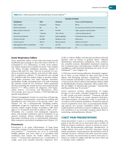
TABLE 22.5 Initial assessment and characteristics of acute asthma 61,62
Severity of attack
Symptoms Mild Moderate Severe or life-threatening
Able to talk in Sentences Phrases Words
Physical exhaustion No No Yes, may have paradoxical chest wall movement
Pulse oximetry (room air) >94% 90–94% <90%; cyanosis may be present
Pulse rate <100/min 100–120/min >120/min; below 60/min
Level of consciousness Normal May be agitated Confused, drowsy or agitated
Wheeze intensity Variable Moderate–loud Often quiet
Central cyanosis Absent May be present Likely to be present
Peak expiratory flow (% predicted) >75% 50–75% <50% or an inability to perform the test
Arterial blood gases Test not necessary If initial response is poor Yes
Acute Respiratory Failure (with or without chills), and mucoid, purulent or bloody
64
Acute respiratory failure occurs when the lungs provide sputum, with an abrupt or gradual onset. Physical
insufficient gas exchange to meet the body’s need for O 2 examination demonstrates tachypnoea, fever, tachycar-
consumption, CO 2 elimination, or both. Acute respira- dia, possible cyanosis, diminished respiratory excursion
63
tory failure results from a number of causes (see Chapter due to pleuritic pain, end-respiratory crackles or rales on
14). When alveolar ventilation decreases, arterial O 2 auscultation with bronchial breathing over areas of con-
64,66
tension falls and CO 2 rises. This rise in arterial CO 2 pro- solidation (see Chapter 13).
duces increased serum carbonic acid and pH falls, result- A CXR may reveal varying infiltrates: interstitial, segmen-
ing in respiratory acidosis. If uncorrected, low arterial tal or lobar; or may initially be clear until later in the
63
O 2 combines with low cardiac output to produce dimin- illness and following adequate rehydration. Venous
55
ished tissue perfusion and tissue hypoxia. Anaerobic blood samples will identify a raised white cell count and/
metabolism results, increasing lactic acid and worsening or leucocytosis. Blood cultures and sputum cultures assist
the acidosis caused by CO 2 retention. Other symptoms in identifying the causative organism. ABGs usually iden-
develop involving the central nervous and cardiovascular tify the degree of impaired gas exchange; hypoxaemia
57
systems. 59,60,63 ABGs confirm the diagnosis, with hyper- and hypocarbia may be present. 66
carbia (PaCO 2 >45 mmHg and hypoxaemia (PaO 2
<80 mmHg), and a low pH evident. A CXR identifies the Initial treatment involves administration of oxygen
specific lung disease. 63 therapy via face-mask, evaluated frequently in response
57
to ABG results and pulse oximetry. Treatment will also
Clinical management focuses on correction of hypercap- require IV fluid therapy to ensure adequate hydration,
nia, treatment of hypoxaemia, correction of acidosis, and and administration of antibiotics orally or parentally in
63
identification and correction of the specific cause (see accordance with antibiotic guidelines. Ventilatory support
Chapter 14). For a spontaneously breathing patient, may be required in some cases; in spontaneously breath-
administer oxygen by ventilation mask (24%) or nasal ing patients non invasive ventilation (NIV) via a face
cannula. Adjust oxygen therapy according to ABG find- mask should be used before invasive ventilation. Mechan-
ings at 15–20-minute intervals to achieve a PaO 2 of 85– ical ventilation is not normally required unless there is
90 mmHg. For a patient with inadequate respiratory underlying cardiopulmonary disease. 57,64,66
effort, non-invasive ventilation may be instituted. In an
apnoeic situation, initiate ventilatory assistance with
bag–mask ventilation prior to endotracheal intubation, CHEST PAIN PRESENTATIONS
then commence mechanical ventilation (see Chapter 15). Chest discomfort or pain is a common presenting com-
plaint to the ED and can be associated with a number of
Pneumonia different clinical conditions, several of which are associ-
Pneumonia is an acute inflammation of lung tissue ated with life-threatening pathology. Identification of
caused by a variety of viral and bacterial organisms, fungi cardiac-related chest pain is therefore important during
and parasites. 64-66 Pneumonia can occur in previously initial assessment, examining pain characteristics such as
healthy patients, but more often it is associated with intensity, location, radiation and other associated symp-
conditions that impair the body’s defence mecha- toms. Consider any presentation in which chest pain is a
nisms. 64,66 Predominant symptoms are a combination of feature as cardiac in origin until this has been ruled out
cough, chest pain (usually pleuritic), dyspnoea, fever or another cause confirmed.