
Page 618 - ACCCN's Critical Care Nursing
P. 618
Emergency Presentations 595
99
l Ischaemic: are precipitated by disrupted blood flow to stroke presentation. IV access is obtained to administer
an area of the brain as a result of arterial occlusion. medications, and collect blood for electrolytes, haemato-
Acute ischaemic stroke presentations are now referred logy and coagulation studies. A blood sugar level test
to as a ‘brain attack’, to promote early presentation for will rule out hypoglycaemia or hyperglycaemia as a cause
access to time-critical treatments, 94,95 and because the of the presenting symptoms. Abnormal glucose levels
pathophysiology and current treatment of acute (isch- adversely affect cerebral metabolism. 99,94 After obvious
aemic) stroke mimics that of acute myocardial infarc- alternative diagnoses are excluded, a brain CT scan deter-
tion (‘heart attack’). From an ED perspective, serious mines whether a stroke is haemorrhagic or ischaemic in
long-term disability can be minimised if ischaemic origin. While a new-onset ischaemic stroke may not be
stroke is recognised and treated promptly; that is, evident for up to 24 hours, blood in the cranial cavity
within 3 hours of symptom onset. 96,97 will be apparent immediately. Patients with any sign of
l Haemorrhagic strokes are caused by rupture of a blood haemorrhage are excluded for fibrinolytic therapy. 95
vessel, which produces bleeding into the brain paren-
chyma. (Chapter 17 details the pathophysiological MANAGEMENT
processes). Acute ischaemic stroke (‘brain attack’) management
For patients diagnosed with a stroke, 30% will die in the includes timely administration of a fibrinolytic agent in
first year after their stroke, most (15–20%) within the first appropriately selected patients (see Box 22.2), which
30 days. Of the 70% who survive, 35% will remain per- facilitates reperfusion, minimises tissue damage and
manently disabled 1 year after a stroke, 10% of whom reduces long-term stroke sequelae. Longer times between
require care in a nursing home or other long-term symptom onset and fibrinolytic infusion are associated
facility. 98,99 with higher rates of morbidity and mortality. 94,98,99,102
Early presentation is therefore essential for appropriate
ASSESSMENT, MONITORING assessments and investigations (including CT scanning)
AND DIAGNOSTICS and thrombolytic administration to fall within the narrow
Symptoms of stroke are a common patient presentation treatment window. This has seen the emergence of acute
stroke units, with specialised teams dedicated to the rapid
to the ED; presenting signs vary from profound altera-
tions in level of consciousness and limb hemiplegia to
mild symptoms affecting speech, cognition or coordina-
tion. Symptoms may include confusion, dizziness, ataxia,
visual disturbances, dysphasia or receptive and expressive
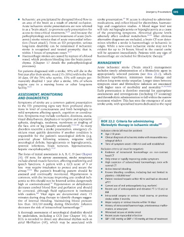
aphasia, dysphagia, weakness, numbness or tingling of BOX 22.2 Criteria for administering
the face, arm or leg (usually unilateral). 97,98,100 As many fibrinolytic therapy in ischaemic stroke 101
disorders resemble a stroke presentation, emergency cli- Inclusion criteria (all must be positive):
nicians must quickly determine if another condition is l Age ≥18 years
responsible for the patient’s neurological deficits (e.g. l Clinical diagnosis of ischaemic stroke with measurable neu-
post-ictal phase following seizures, migraine with rological deficit
neurological deficits, hypoglycaemia or hyperglycaemia, l Time of symptom onset <180 min and well established
systemic infections, brain tumours, hyponatraemia,
hepatic encephalopathy). 93,97 Exclusion criteria (all must be negative):
l Evidence of intracranial haemorrhage on non-contrast
The focus of initial assessment is A, B, C, D (see Chapter head CT
24). Of note, for airway assessment, stroke symptoms l Only minor or rapidly improving stroke symptoms
include altered muscle function, affecting swallowing and l High suspicion of subarachnoid haemorrhage, even with
speech functions. A patient with a GCS score of 9 or normal CT
less may require intubation to protect and secure the l Active internal bleeding
airway. 99,101 The patient’s breathing pattern should be l Known bleeding condition, including but not limited to
assessed and continually monitored. Hypertension is platelets <100,000/mm 3
common, with the increase improving any cerebral isch- l Patient received heparin within 48 hr and had an elevated
aemia so this should not be lowered unless dangerously aPTT
94
high or contraindicated. Hypotension or dehydration l Current use of oral anticoagulants (e.g. warfarin)
decreases cerebral blood flow and perfusion and should l Recent use of anticoagulant and elevated PT (>15 sec) or
be corrected, although fluid replacement is instituted INR
100
with caution. Vital signs are documented every 15 l Intracranial surgery or serious head trauma, or previous
minutes during drug therapy to identify changes sugges- stroke within 3 months
tive of internal bleeding. Maintaining blood pressure l Major surgery or serious trauma within 14 days
less than 185/110 mmHg during fibrinolytic infusion l History of intracranial haemorrhage, arteriovenous malfor-
decreases the risk of intracerebral haemorrhage. 95
mation, or aneurysm
A thorough assessment of neurological disability should l Witnessed seizure at stroke onset
be undertaken, including a GCS (see Chapter 16). An l Recent acute myocardial infarction
ECG is recorded to detect any abnormal rhythm such as l SBP >185 mmHg or DBP >110 mmHg at time of treatment
atrial fibrillation (AF), which may be associated with