
Page 650 - ACCCN's Critical Care Nursing
P. 650
Trauma Management 627
Cervical Spine Immobilisation Procedure
Cervical spine immobilisation should be performed as a team. Generally, four people should work together.
1. Leader is positioned at the head of the patient and positions his or her hands on each side of the patient’s head, with thumbs along
the mandible and fingers behind the head on the occipital ridge. Maintain gentle but firm stabilization of the patient’s neck
throughout the entire procedure.
2. Assess the patient’s motor and sensory level by asking the patient to wiggle his or her toes and fingers. Touch the patient’s arms
and legs to determine sensory response.
3. Apply and secure appropriate fitting cervical collar. Follow the directions for sizing that comes with each collar. An ill-fitting collar
can cause pain, occlude the patient’s airway, or fail to give appropriate immobilisation.
4. Straighten the patient’s arms and legs and position team members so that the patient may be rolled on the backboard as a unit.
5. The patient’s head should be immobilised until the straps are correctly placed. The straps should be placed so that the patient is secured
to the backboard at the shoulders, hips, and proximal to the knees.
6. The patient’s head should be further immobilised with head blocks or towel rolls. Tape or straps should not be placed across the chin.
7. The patient’s head should be manually immobilised until the head and neck are immobilised.
8. The patient’s motor and sensory function should be reassessed after the patient is immobilised.
9. Some patients such as those with a compromised airway or neck deformities may not be able to tolerate laying flat.
10. Massive neck swelling that may result from a penetrating injury may prohibit the use of a cervical collar. Towel rolls and tape may
be safer method of securing the patient to the board and allow for evaluation of the patient’s injury.
Modified from Emergency Nurses Association: Trauma nursing core course provider manual, ed 5, Des Plaines, III, 2000, The Association.
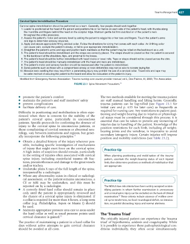
78
FIGURE 23.1 Spine Movement Precautions.
l promote the patient’s comfort The two methods available for moving the trauma patient
l maintain the patient’s and staff members’ safety are staff manual handling and lifting hoists. Generally,
l prevent complications trauma patients can be log-rolled (see Figure 23.1 for
l facilitate delivery of care. initial care and p. 635 for later care) as frequently as
required for nursing care. Any restrictions to patient posi-
Difficulty in positioning and mobilisation is often expe- tioning and weight bearing due to injuries or physiologi-
rienced when there is concern for the stability of the cal status must be considered through this process; it is
patient’s cervical spine, particularly in unconscious essential that care be taken to prevent any worsening of
patients. Specific protocols for confirming the absence of injuries due to handling of the patient. Knowledge of the
injury to the cervical spine in unconscious patients, or position restrictions for each limb, including all weight-
those complaining of cervical soreness or abnormal neu- bearing joints and the vertebrae, is imperative to avoid
rology, vary between institutions and regions, but gener- secondary iatrogenic injury. Certain injuries will impose
ally incorporate the following principles: 31
position and mobility restrictions (see Table 23.2).
l Obtain a detailed history of the injury wherever pos-
sible, including specific investigation of mechanisms
of injury that might exert force on the cervical spine. Practice tip
A high index of suspicion should remain, particularly
in the setting of injuries often associated with cervical When planning positioning and mobilisation of the trauma
spine injury, including craniofacial trauma rib frac- patient, ascertain the weight-bearing status of each injured
tures, pneumothoraces and damage to the great vessels limb, then determine positions or methods of mobilisation that
and/or trachea. are appropriate.
l Undertake plain X-rays of the full length of the spine,
interpreted by a radiologist.
l Where any abnormality exists in clinical or radiologi-
cal assessment, or the patient remains unconscious, a Practice tip
CT or MRI may be undertaken, and this must be
reported on by a radiologist. The NEXUS low-risk criteria have been widely accepted as iden-
l A correctly fitted hard collar should remain in place tifying patients in whom further examination is unnecessary
only until the patient is appropriately reviewed and and cervical spine injury can be excluded on the basis of clinical
the chance of a cervical spine injury is eliminated. If examination. These criteria include absence of midline cervi-
82
a collar is required for more than 4 hours, a long-term cal spine tenderness, no focal neurological deficit, no intoxica-
collar (e.g. Philadelphia, Aspen or Miami J) should tion, no painful distracting injury and normal alertness.
be used.
l Maintain appropriate pressure area care to areas under
the hard collar as well as usual pressure points until The ‘Trauma Triad’
cervical clearance is gained. 32
The critically injured patient can experience the ‘trauma
The practice of maintaining a patient in a hard collar for triad’ of hypothermia, acidosis and coagulopathy. While
days without active attempts to gain cervical clearance it is possible to experience these pathophysiological con-
should be avoided at all costs. ditions individually, they often occur simultaneously.