
Page 81 - ACCCN's Critical Care Nursing
P. 81
58 S C O P E O F C R I T I C A L C A R E
recovering from Acute Lung Injury/Adult Respiratory Dis-
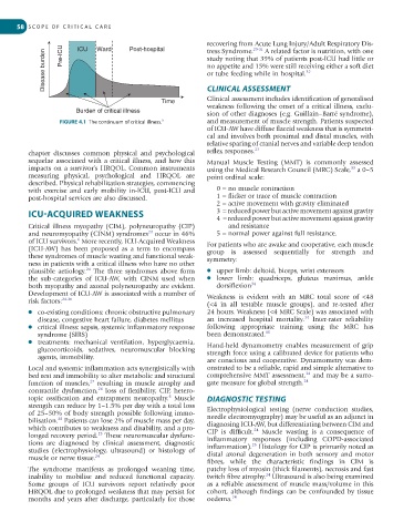
Pre-ICU
Post-hospital
ICU
Ward
29-31
A related factor is nutrition, with one
tress Syndrome.
Disease burden study noting that 39% of patients post-ICU had little or
no appetite and 15% were still receiving either a soft diet
32
or tube feeding while in hospital.
Clinical assessment includes identification of generalised
Time CLINICAL ASSESSMENT
weakness following the onset of a critical illness, exclu-
Burden of critical illness sion of other diagnoses (e.g. Guillain–Barré syndrome),
FIGURE 4.1 The continuum of critical illness. 9 and measurement of muscle strength. Patients suspected
of ICU-AW have diffuse flaccid weakness that is symmetri-
cal and involves both proximal and distal muscles, with
relative sparing of cranial nerves and variable deep tendon
chapter discusses common physical and psychological reflex responses. 23
sequelae associated with a critical illness, and how this Manual Muscle Testing (MMT) is commonly assessed
impacts on a survivor’s HRQOL. Common instruments using the Medical Research Council (MRC) Scale, a 0–5
33
measuring physical, psychological and HRQOL are point ordinal scale:
described. Physical rehabilitation strategies, commencing
with exercise and early mobility in-ICU, post-ICU and 0 = no muscle contraction
post-hospital services are also discussed. 1 = flicker or trace of muscle contraction
2 = active movement with gravity eliminated
ICU-ACQUIRED WEAKNESS 3 = reduced power but active movement against gravity
4 = reduced power but active movement against gravity
Critical illness myopathy (CIM), polyneuropathy (CIP) and resistance
23
and neuromyopathy (CINM) syndromes occur in 46% 5 = normal power against full resistance.
of ICU survivors. More recently, ICU-Acquired Weakness For patients who are awake and cooperative, each muscle
6
(ICU-AW) has been proposed as a term to encompass group is assessed sequentially for strength and
these syndromes of muscle wasting and functional weak- symmetry:
ness in patients with a critical illness who have no other
24
plausible aetiology. The three syndromes above form ● upper limb: deltoid, biceps, wrist extensors
the sub-categories of ICU-AW, with CINM used when ● lower limb: quadriceps, gluteus maximus, ankle
both myopathy and axonal polyneuropathy are evident. dorsiflexion 34
Development of ICU-AW is associated with a number of Weakness is evident with an MRC total score of <48
risk factors: 24-26
(<4 in all testable muscle groups), and re-tested after
● co-existing conditions: chronic obstructive pulmonary 24 hours. Weakness (<4 MRC Scale) was associated with
34
disease, congestive heart failure, diabetes mellitus an increased hospital mortality. Inter-rater reliability
● critical illness: sepsis, systemic inflammatory response following appropriate training using the MRC has
syndrome (SIRS) been demonstrated. 35
● treatments: mechanical ventilation, hyperglycaemia, Hand-held dynamometry enables measurement of grip
glucocorticoids, sedatives, neuromuscular blocking strength force using a calibrated device for patients who
agents, immobility. are conscious and cooperative. Dynamometry was dem-
Local and systemic inflammation acts synergistically with onstrated to be a reliable, rapid and simple alternative to
34
bed rest and immobility to alter metabolic and structural comprehensive MMT assessment, and may be a surro-
27
function of muscles, resulting in muscle atrophy and gate measure for global strength. 24
26
contractile dysfunction, loss of flexibility, CIP, hetero-
topic ossification and entrapment neuropathy. Muscle DIAGNOSTIC TESTING
6
strength can reduce by 1–1.5% per day with a total loss
of 25–50% of body strength possible following immo- Electrophysiological testing (nerve conduction studies,
bilisation. Patients can lose 2% of muscle mass per day, needle electromyography) may be useful as an adjunct in
28
which contributes to weakness and disability, and a pro- diagnosing ICU-AW, but differentiating between CIM and
24
25
longed recovery period. These neuromuscular dysfunc- CIP is difficult. Muscle wasting is a consequence of
tions are diagnosed by clinical assessment, diagnostic inflammatory responses (including COPD-associated
25
studies (electrophysiology, ultrasound) or histology of inflammation). Histology for CIP is primarily noted as
muscle or nerve tissue. 24 distal axonal degeneration in both sensory and motor
fibres, while the characteristic findings in CIM is
The syndrome manifests as prolonged weaning time, patchy loss of myosin (thick filaments), necrosis and fast
24
inability to mobilise and reduced functional capacity. twitch fibre atrophy. Ultrasound is also being examined
Some groups of ICU survivors report relatively poor as a reliable assessment of muscle mass/volume in this
HRQOL due to prolonged weakness that may persist for cohort, although findings can be confounded by tissue
months and years after discharge, particularly for those oedema. 24