
Page 223 - Concise Pathology for Exam Preparation ( PDFDrive )
P. 223
208 SECTION I General Pathology
2. Nonimmune hydrops
• Major causes include cardiovascular defects, chromosomal anomalies (Turner
syndrome, trisomies 18 and 21) and fetal anaemia (eg, that associated with
homozygous a-thalassaemia) resulting in intrauterine cardiac failure.
• Transplacental infection with parvovirus B19 is emerging as an important cause of
fetal hydrops.
Morphology of Hydrops
- Presence of dysmorphic features suggests underlying chromosomal abnormalities.
- Postmortem examination may reveal a cardiac anomaly.
- In hydrops associated with fetal anaemia, both the fetus and the placenta are character-
istically pale.
- In most cases, there is hepatosplenomegaly.
- Compensatory erythroid hyperplasia may be seen in the marrow and extramedullary hae-
matopoiesis may be seen in liver, spleen, kidneys and lungs.
- Haemolysis leads to increased unconjugated bilirubin.
- CNS is damaged when bilirubin levels are more than 20 mg/dL. Basal ganglia and brain
stem are prone to deposition of bilirubin (Kernicterus).
Q. Enumerate the common malignant tumours of infancy
and childhood.
Ans. Common malignant tumours of infancy and childhood are enlisted in Table 8.3.
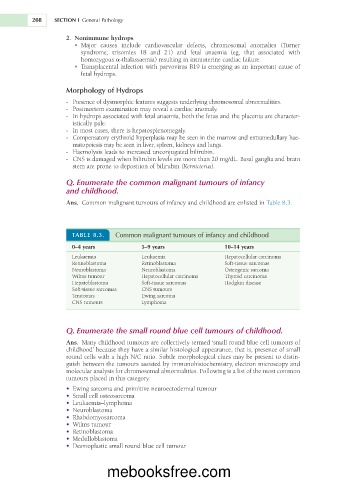
TABLE 8.3. Common malignant tumours of infancy and childhood
0–4 years 5–9 years 10–14 years
Leukaemia Leukaemia Hepatocellular carcinoma
Retinoblastoma Retinoblastoma Soft-tissue sarcomas
Neuroblastoma Neuroblastoma Osteogenic sarcoma
Wilms tumour Hepatocellular carcinoma Thyroid carcinoma
Hepatoblastoma Soft-tissue sarcomas Hodgkin disease
Soft-tissue sarcomas CNS tumours
Teratomas Ewing sarcoma
CNS tumours Lymphoma
Q. Enumerate the small round blue cell tumours of childhood.
Ans. Many childhood tumours are collectively termed ‘small round blue cell tumours of
childhood’ because they have a similar histological appearance, that is, presence of small
round cells with a high N/C ratio. Subtle morphological clues may be present to distin-
guish between the tumours assisted by immunohistochemistry, electron microscopy and
molecular analysis for chromosomal abnormalities. Following is a list of the most common
tumours placed in this category:
• Ewing sarcoma and primitive neuroectodermal tumour
• Small cell osteosarcoma
• Leukaemia–lymphoma
• Neuroblastoma
• Rhabdomyosarcoma
• Wilms tumour
• Retinoblastoma
• Medulloblastoma
• Desmoplastic small round blue cell tumour
mebooksfree.com