
Page 327 - Concise Pathology for Exam Preparation ( PDFDrive )
P. 327
312 SECTION II Diseases of Organ Systems
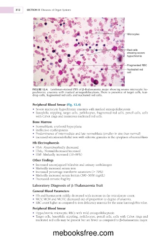
Microcytes
Red cells
showing severe
hypochromia
Fragmented RBC
Nucleated red
cell
FIGURE 12.4. Leishman-stained PBS of b-thalassaemia major showing severe microcytic hy-
pochromic anaemia with marked anisopoikilocytosis. There is presence of target cells, tear-
drop cells, fragmented red cells and nucleated red cells.
Peripheral Blood Smear (Fig. 12.4)
• Severe microcytic hypochromic anaemia with marked anisopoikilocytosis
• Basophilic stippling, target cells, poikilocytes, fragmented red cells, pencil cells, cells
with Cabot rings and numerous nucleated red cells.
Bone Marrow
• Normoblastic erythroid hyperplasia
• Ineffective erythropoiesis
• Predominance of intermediate and late normoblasts (smaller in size than normal)
• Increased reticuloendothelial iron with siderotic granules in the cytoplasm of normoblasts
Hb Electrophoresis
• HbA: Absent/markedly decreased
• HbA 2 : Normal/decreased/increased
• HbF: Markedly increased (10–98%)
Other Findings
• Increased unconjugated bilirubin and urinary urobilinogen
• Markedly increased serum iron
• Increased percentage transferrin saturation (. 70%)
• Markedly increased serum ferritin (300–3000 mg/dL)
• Decreased osmotic fragility
Laboratory Diagnosis of b-Thalassaemia Trait
General Blood Parameters
• Hb and haematocrit mildly decreased with increase in the reticulocyte count.
• MCV, MCH and MCHC decreased out of proportion to degree of anaemia.
• RBC count higher as compared to iron deficiency anaemia for the same haemoglobin value.
Peripheral Blood Smear
• Hypochromic microcytic RBCs with mild anisopoikilocytosis
• Target cells, basophilic stippling, poikilocytes, pencil cells, cells with Cabot rings and
nucleated red cells may be present but are fewer as compared to b-thalassaemia major.
mebooksfree.com