
Page 326 - Concise Pathology for Exam Preparation ( PDFDrive )
P. 326
12 Haematology 311
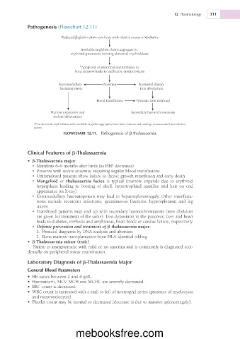
Pathogenesis (Flowchart 12.11)
Reduced β-globin chain synthesis with relative excess of α-chains
Insoluble α-globin chains aggregate in
erythroid precursors forming abnormal erythroblasts
*Apoptosis of abnormal erythroblasts in
bone marrow leads to ineffective erythropoiesis
Extramedullary Anaemia Increased dietary
haematopoiesis iron absorption
Blood transfusion Systemic iron overload
Marrow expansion and Secondary haemochromatosis
skeletal deformities
*Few abnormal erythroblasts with insoluble α-globin aggregates leave bone marrow and undergo extravascular haemolysis in
spleen.
FLOWCHART 12.11. Pathogenesis of b-thalassaemia.
Clinical Features of b-Thalassaemia
• b-Thalassaemia major
• Manifests 6–9 months after birth (as HbF decreases)
• Presents with severe anaemia, requiring regular blood transfusions
• Untransfused patients show failure to thrive; growth retardation and early death
• Mongoloid or thalassaemia facies is typical (marrow expands due to erythroid
hyperplasia leading to bossing of skull, hypertrophied maxillae and hair on end
appearance on X-ray).
• Extramedullary haematopoiesis may lead to hepatosplenomegaly. Other manifesta-
tions include recurrent infections, spontaneous fractures, hypersplenism and leg
ulcers.
• Transfused patients may end up with secondary haemochromatosis (iron chelators
are given for treatment of the same). Iron deposition in the pancreas, liver and heart
leads to diabetes, cirrhosis and arryhtmias, heart block or cardiac failure, respectively.
• Definite prevention and treatment of b-thalassaemia major:
1. Prenatal diagnosis by DNA analysis and abortion
2. Bone marrow transplantation from HLA-identical sibling
• b-Thalassaemia minor (trait)
Patient is asymptomatic with mild or no anaemia and is commonly is diagnosed acci-
dentally on peripheral smear examination.
Laboratory Diagnosis of b-Thalassaemia Major
General Blood Parameters
• Hb varies between 2 and 6 g/dL.
• Haematocrit, MCV, MCH and MCHC are severely decreased.
• RBC count is decreased.
• WBC count is increased with a shift to left of neutrophil series (presence of myelocytes
and metamyelocytes).
• Platelet count may be normal or decreased (decrease is due to massive splenomegaly).
mebooksfree.com