
Page 322 - Concise Pathology for Exam Preparation ( PDFDrive )
P. 322
12 Haematology 307
G6PD
Glucose-6-phosphate 6-phosphogluconate
2GSH NADP
NADPH
Glutathione Glutathione
peroxidase reductase
GSSG
H O H O
2 2
2
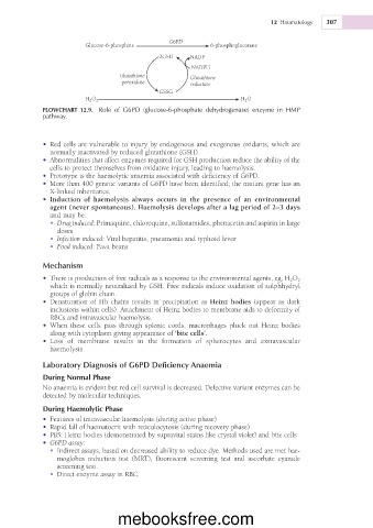
FLOWCHART 12.9. Role of G6PD (glucose-6-phosphate dehydrogenase) enzyme in HMP
pathway.
• Red cells are vulnerable to injury by endogenous and exogenous oxidants, which are
normally inactivated by reduced glutathione (GSH).
• Abnormalities that affect enzymes required for GSH production reduce the ability of the
cells to protect themselves from oxidative injury, leading to haemolysis.
• Prototype is the haemolytic anaemia associated with deficiency of G6PD.
• More than 400 genetic variants of G6PD have been identified; the mutant gene has an
X-linked inheritance.
• Induction of haemolysis always occurs in the presence of an environmental
agent (never spontaneous). Haemolysis develops after a lag period of 2–3 days
and may be:
• Drug induced: Primaquine, chloroquine, sulfonamides, phenacetin and aspirin in large
doses
• Infection induced: Viral hepatitis, pneumonia and typhoid fever
• Food induced: Fava beans
Mechanism
• There is production of free radicals as a response to the environmental agents, eg, H 2 O 2
which is normally neutralized by GSH. Free radicals induce oxidation of sulphhydryl
groups of globin chain.
• Denaturation of Hb chains results in precipitation as Heinz bodies (appear as dark
inclusions within cells). Attachment of Heinz bodies to membrane aids to deformity of
RBCs and intravascular haemolysis.
• When these cells pass through splenic cords, macrophages pluck out Heinz bodies
along with cytoplasm giving appearance of ‘bite cells’.
• Loss of membrane results in the formation of spherocytes and extravascular
haemolysis.
Laboratory Diagnosis of G6PD Deficiency Anaemia
During Normal Phase
No anaemia is evident but red cell survival is decreased. Defective variant enzymes can be
detected by molecular techniques.
During Haemolytic Phase
• Features of intravascular haemolysis (during active phase)
• Rapid fall of haematocrit with reticulocytosis (during recovery phase)
• PBS: Heinz bodies (demonstrated by supravital stains like crystal violet) and bite cells
• G6PD assay:
• Indirect assays, based on decreased ability to reduce dye. Methods used are met hae-
moglobin reduction test (MRT), fluorescent screening test and ascorbate cyanide
screening test.
• Direct enzyme assay in RBC.
mebooksfree.com