
Page 324 - Concise Pathology for Exam Preparation ( PDFDrive )
P. 324
12 Haematology 309
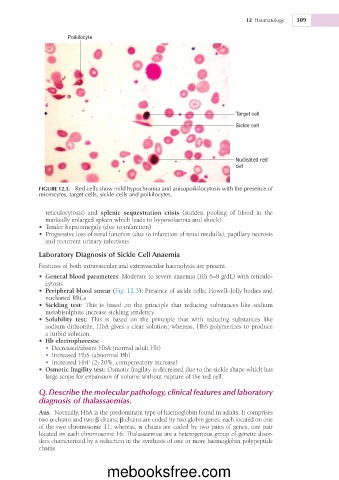
Poikilocyte
Target cell
Sickle cell
Nucleated red
cell
FIGURE 12.3. Red cells show mild hypochromia and anisopoikilocytosis with the presence of
microcytes, target cells, sickle cells and poikilocytes.
reticulocytosis) and splenic sequestration crisis (sudden pooling of blood in the
markedly enlarged spleen which leads to hypovolaemia and shock).
• Tender hepatomegaly (due to infarction)
• Progressive loss of renal function (due to infarction of renal medulla), papillary necrosis
and recurrent urinary infections
Laboratory Diagnosis of Sickle Cell Anaemia
Features of both intravascular and extravascular haemolysis are present
• General blood parameters: Moderate to severe anaemia (Hb 6–8 g/dL) with reticulo-
cytosis
• Peripheral blood smear (Fig. 12.3): Presence of sickle cells; Howell–Jolly bodies and
nucleated RBCs
• Sickling test: This is based on the principle that reducing substances like sodium
metabisulphite increase sickling tendency.
• Solubility test: This is based on the principle that with reducing substances like
sodium dithionite, HbA gives a clear solution; whereas, HbS polymerizes to produce
a turbid solution.
• Hb electrophoresis:
• Decreased/absent HbA (normal adult Hb)
• Increased HbS (abnormal Hb)
• Increased HbF (2–20%, compensatory increase)
• Osmotic fragility test: Osmotic fragility is decreased due to the sickle shape which has
large scope for expansion of volume without rupture of the red cell.
Q. Describe the molecular pathology, clinical features and laboratory
diagnosis of thalassaemias.
Ans. Normally, HbA is the predominant type of haemoglobin found in adults. It comprises
two a chains and two b chains; b chains are coded by two globin genes, each located on one
of the two chromosome 11; whereas, a chains are coded by two pairs of genes, one pair
located on each chromosome 16. Thalassaemias are a heterogenous group of genetic disor-
ders characterized by a reduction in the synthesis of one or more haemoglobin polypeptide
chains.
mebooksfree.com