
Page 334 - Concise Pathology for Exam Preparation ( PDFDrive )
P. 334
12 Haematology 319
2. Lymphoid leukemoid reactions
• Infections like infectious mononucleosis, cytomegalovirus, pertussis, mumps, measles,
rubella, tuberculosis, syphilis, brucellosis and infective hepatitis
• CLL
• Carcinoma
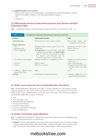
Q. Differentiate between leukemoid reactions and chromic myeloid
leukaemia (CML).
Ans. Comparative features of leukemoid reactions and CML are tabulated in Table 12.9.
TABLE 12.9. Comparison between leukaemoid reactions and CML
Features Leukaemoid reaction CML
Clinical features Clinical features of the causative disorder Splenomegaly, lymph node
enlargement and anaemia
Blood examination
9
9
• TLC Moderate increase; rarely exceeds 100 3 10 /L Usual range 20–500 3 10 /L
• Immature cells Usually few Usually numerous
• WBC morphology Toxic granules (increased number of intensely Uncommon
staining primary granules) and Dohle bodies
(discrete round to oval cytoplasmic bodies,
1–2 microns, stain blue-grey with Ro-
manowski stains) seen in infective cases
• Eosinophilia and basophilia Absolute eosinophilia or basophilia not seen Absolute eosinophilia or baso-
philia seen
• Anaemia Slight or absent Present and progressive
• Platelets Normal or increased Increased; may decrease in ac-
celerated phase and blast
crisis
• Leukocyte alkaline phos- High Low
phatase (LAP) score
Autopsy No infiltration of organs and tissues Leukaemic infiltration of or-
gans and tissues is present
Q. Define and enumerate the myeloproliferative disorders?
Ans. Myeloproliferative disorders occur due to clonal expansion of a multipotent haema-
topoietic progenitor cells with the overproduction of one or more of the formed elements
of the blood. These conditions may evolve into acute leukaemia. The following conditions
are included under this category of diseases:
• CML
• Polycythaemia vera
• Essential thrombocythaemia
• Primary myelofibrosis
• Systemic mastocytosis
• Chronic eosinophilic leukaemia
• Stem cell leukaemia
Q. Define and classify polycythaemia?
Ans. It is defined as neoplastic proliferation of erythroid, granulocytic and megakaryocytic
elements. Polycythaemia can be classified as:
1. Relative: Relative polycythemia results from haemoconcentration due to reduced plasma
volume (seen in dehydration—low fluid intake, vomiting, diaorrhea and excessive sweat-
ing). The red cell mass remains within the normal range in relative polycythemia.
mebooksfree.com