
Page 337 - Concise Pathology for Exam Preparation ( PDFDrive )
P. 337
322 SECTION II Diseases of Organ Systems
TABLE 12.11. FAB classification of acute myeloid leukaemias (AML)—cont’d
• M4Eo Variant Shows increase in marrow eosinophils
M5 Monocytic leukaemia More than 80% cells in the bone marrow are monocytic (monoblasts,
(a) Undifferentiated (mono- promonocytes and monocytes)
blastic) M5a In M5a, 80% or more cells are monoblasts
(b) Well-differentiated In M5b, predominant cells are promonocytes and monocytes
(promonocytic-mono- Variable expression of myeloid antigens CD33, CD13 and CD117
cytic) M5b Monocytic markers CD14, CD36, CD64 and CD11c are positive
M6 Erythroleukaemia Predominance of erythroblasts. It has two subtypes: Erythroleukae-
(Di Guglielmo disease) mia (. 20% of nonerythroid cells are myeloblasts and . 50% of
all nucleated cells are erythroblasts) and pure erythroid leukaemia
(. 80% of marrow cells are erythroblasts). Erythroblasts may be
bizarre looking with bi- and trinucleate forms and megaloblastic
nuclear features and are positive for monoclonal antibody against
glycophorin A
M7 Megakaryoblastic leukaemia Blasts are more than 20% of which at least 50% are of megakaryocytic
origin. Megakaryoblasts resemble lymphoblasts but show distinct
cytoplasmic blebs or pseudopod formation. Cytochemically, they
are negative for myeloperoxidase and positive for platelet peroxi-
dase. Megakaryoblasts express CD41 (glycoprotein IIb/IIIa) and/or
CD61 (glycoprotein IIIa)
TABLE 12.12. FAB classification of acute lymphoid leukaemias (ALL)
L1 85% Morphology: L1 blasts are small and homogeneous. The nuclei are round and regular
with little clefting and inconspicuous nucleoli. Cytoplasm is lightly basophilic, scanty
and usually without vacuoles
Staining: MPO is always negative
Maturation: pro-B or pre-B lineage
L2 14% Morphology: L2 blasts are large and heterogeneous. The nuclei are irregular and often
clefted. One or more, usually large nucleoli are present. The volume of cytoplasm is
variable, but often abundant and may contain vacuoles
Cytochemistry: L2 blasts may have granular PAS positivity. MPO is negative
Maturation: pro-B or pre-B and T-cell ALL lineage
L3 Burkitt’s 1% Morphology: L3 blasts are large in size and homogeneous. The nuclei are regular and
round-oval in shape. One or more prominent nucleoli are present. They have moderate
to abundant deeply basophilic cytoplasm, which contains prominent vacuoles
Cytochemistry: MPO is always negative. NSE is usually negative, but may show focal
cytoplasmic positivity. Vacuoles are PAS-negative but are classically positive for the
neutral lipid stain Oil Red O
Maturation: All L3 leukaemias are surface immunoglobulin (SIg)-positive and are of
B-cell lineage
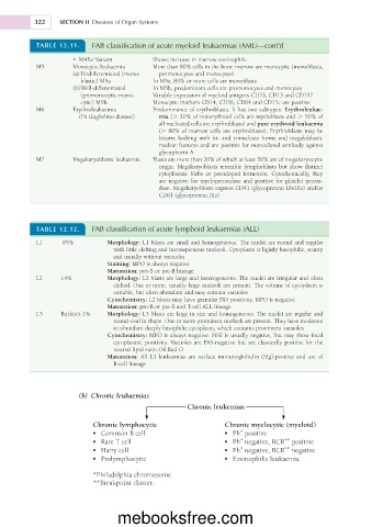
(b) Chronic leukaemias
Chronic leukemias
Chronic lymphocytic Chronic myelocytic (myeloid)
• Common B cell • Ph positive
*
*
**
• Rare T cell • Ph negative, BCR positive
**
• Hairy cell • Ph negative, BCR negative
*
• Prolymphocytic • Eosinophilic leukaemia
*Philadelphia chromosome.
**Breakpoint cluster.
mebooksfree.com