
Page 336 - Concise Pathology for Exam Preparation ( PDFDrive )
P. 336
12 Haematology 321
Q. Differentiate between primary and secondary polycythaemia.
Ans. Contrasting features of primary and secondary polycythaemia are tabulated in Table 12.10.
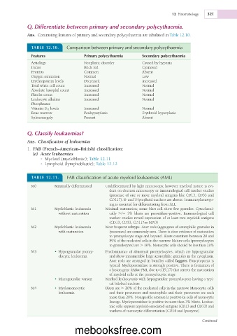
TABLE 12.10. Comparison between primary and secondary polycythaemia
Features Primary polycythaemia Secondary polycythaemia
Aetiology Neoplastic disorder Caused by hypoxia
Facies Brick red Cyanosed
Pruritus Common Absent
Oxygen saturation Normal Low
Erythropoietin levels Decreased Increased
Total white cell count Increased Normal
Absolute basophil count Increased Normal
Platelet count Increased Normal
Leukocyte alkaline Increased Normal
Phosphatase
Vitamin B 12 levels Increased Normal
Bone marrow Panhyperplasia Erythroid hyperplasia
Splenomegaly Present Absent
Q. Classify leukaemias?
Ans. Classification of leukaemias
1. FAB (French–American–British) classification:
(a) Acute leukaemias
• Myeloid (myeloblastic); Table 12.11
• Lymphoid (lymphoblastic); Table 12.12
TABLE 12.11. FAB classification of acute myeloid leukaemias (AML)
M0 Minimally differentiated Undifferentiated by light microscopy, however myeloid nature is evi-
dent on electron microscopy or immunological cell marker studies
(presence of one or more myeloid antigens like CD13, CD33 and
CD117). B- and T-lymphoid markers are absent. Immunophenotyp-
ing is essential for differentiating from ALL
M1 Myeloblastic leukaemia Minimal maturation; some blast cell show few granules. Cytochemi-
without maturation cally ./5 3% blasts are peroxidase-positive. Immunological cell
marker studies reveal expression of at least two myeloid antigens
(CD13, CD33, CD117or MPO)
M2 Myeloblastic leukaemia Most frequent subtype. Auer rods (aggregates of azurophilic granules in
with maturation lysosomes) are commonly seen. There is clear evidence of maturation
to promyelocyte stage and beyond. Blasts constitute between 20 and
89% of the nucleated cells in the marrow. Mature cells (promyelocytes
to granulocytes) are . 10%. Monocytic cells should be less than 20%
M3 • Hypergranular promy- Predominance of abnormal promyelocytes, which are hypergranular
elocytic leukaemia and show innumerable large azurophilic granules in the cytoplasm.
Auer rods are arranged in bundles called faggots. Pancytopenia is
typical. Myeloperoxidase is strongly positive. There is formation of
a fusion gene RARa-PML due to t(15;17) that arrests the maturation
of myeloid cells at the promyelocytic stage
• Microgranular variant Marked leukocytosis with hypogranular promyelocytes having a typi-
cal bilobed nucleus
M4 • Myelomonocytic Blasts are . 20% of the nucleated cells in the marrow. Monocytic cells
leukaemia and their precursors and neutrophils and their precursors are each
more than 20%. Nonspecific esterase is positive in cells of monocytic
lineage. Myeloperoxidase is positive in more than 3% blasts. Leukae-
mic cells express myeloid-associated antigens (CD13 and CD33) and
markers of monocytic differentiation (CD14 and lysozyme)
Continued
mebooksfree.com