
Page 338 - Concise Pathology for Exam Preparation ( PDFDrive )
P. 338
12 Haematology 323
2. World Health Organization (WHO) classification of acute leukaemias
Blast count for diagnosis of acute leukaemia . or 5 20% in peripheral blood or bone
marrow (in FAB classification the cut off is 30%; it has been demonstrated that
the survival pattern of patients with 20–30% blasts is similar to those with a count
of .30%).
WHO classification of AML
• AML with recurrent genetic abnormalities
• AML with t(8; 21) (q22; q22); AML1/ETO
• AML with abnormal bone marrow eosinophils inv(16)(p13; q22) or t(16; 16)(p13;
q22); (CBFb/MYH11)
• Acute promyelocytic leukaemia AML with t(15; 17)(q22; q12)(PML/RARa) and variants
• AML with 11q23(MLL) abnormalities
• AML with multilineage dysplasia
• Following a myelodysplastic syndrome
• Without antecedent myelodysplastic syndrome
• AML and myelodysplastic syndromes, therapy related
• Alkylating agent related
• Topoisomerase Type II inhibitor related
• Other types
• AML not otherwise characterized/specified
• AML minimally differentiated
• AML without maturation
• AML with maturation
• Acute myelomonocytic leukaemia
• Acute monoblastic and monocytic leukaemia
• Acute erythroid leukaemia
• Acute megakaryoblastic leukaemia
• Acute basophilic leukaemia
• Acute pan myelosis with myelofibrosis
• Myeloid sarcoma
• Myeloid proliferations related to Down’s syndrome
• Blastic plasmacytoid dendritic cell neoplasms
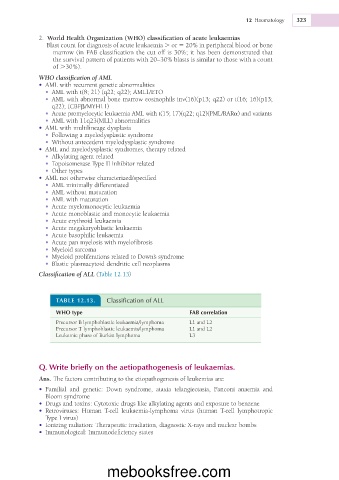
Classification of ALL (Table 12.13)
TABLE 12.13. Classification of ALL
WHO type FAB correlation
Precursor B lymphoblastic leukaemia/lymphoma L1 and L2
Precursor T lymphoblastic leukaemia/lymphoma L1 and L2
Leukemic phase of Burkitt lymphoma L3
Q. Write briefly on the aetiopathogenesis of leukaemias.
Ans. The factors contributing to the etiopathogenesis of leukemias are:
• Familial and genetic: Down syndrome, ataxia telangiectasia, Fanconi anaemia and
Bloom syndrome
• Drugs and toxins: Cytotoxic drugs like alkylating agents and exposure to benzene
• Retroviruses: Human T-cell leukaemia-lymphoma virus (human T-cell lymphotropic
Type I virus)
• Ionizing radiation: Therapeutic irradiation, diagnostic X-rays and nuclear bombs
• Immunological: Immunodeficiency states
mebooksfree.com