
Page 390 - Concise Pathology for Exam Preparation ( PDFDrive )
P. 390
13 The Lung 375
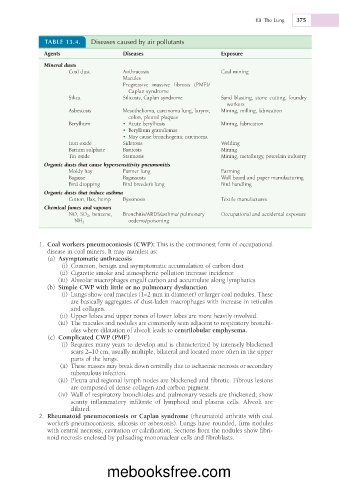
TABLE 13.4. Diseases caused by air pollutants
Agents Diseases Exposure
Mineral dusts
Coal dust Anthracosis Coal mining
Macules
Progressive massive fibrosis (PMF)/
Caplan syndrome
Silica Silicosis, Caplan syndrome Sand blasting, stone cutting, foundry
workers
Asbestosis Mesothelioma, carcinoma lung, larynx, Mining, milling, fabrication
colon, pleural plaques
Beryllium • Acute berylliosis Mining, fabrication
• Beryllium granulomas
• May cause bronchogenic carcinoma
Iron oxide Siderosis Welding
Barium sulphate Baritosis Mining
Tin oxide Stannosis Mining, metallurgy, porcelain industry
Organic dusts that cause hypersensitivity pneumonitis
Moldy hay Farmer lung Farming
Bagasse Bagassosis Wall board and paper manufacturing
Bird dropping Bird breeder’s lung Bird handling
Organic dusts that induce asthma
Cotton, flax, hemp Byssinosis Textile manufactures
Chemical fumes and vapours
NO, SO 2 , benzene, Bronchitis/ARDS/asthma/ pulmonary Occupational and accidental exposure
NH 3 oedema/poisoning
1. Coal workers pneumoconiosis (CWP): This is the commonest form of occupational
disease in coal miners. It may manifest as:
(a) Asymptomatic anthracosis
(i) Common, benign and asymptomatic accumulation of carbon dust
(ii) Cigarette smoke and atmospheric pollution increase incidence
(iii) Alveolar macrophages engulf carbon and accumulate along lymphatics
(b) Simple CWP with little or no pulmonary dysfunction
(i) Lungs show coal macules (1–2 mm in diameter) or larger coal nodules. These
are basically aggregates of dust-laden macrophages with increase in reticulin
and collagen.
(ii) Upper lobes and upper zones of lower lobes are more heavily involved.
(iii) The macules and nodules are commonly seen adjacent to respiratory bronchi-
oles where dilatation of alveoli leads to centrilobular emphysema.
(c) Complicated CWP (PMF)
(i) Requires many years to develop and is characterized by intensely blackened
scars 2–10 cm, usually multiple, bilateral and located more often in the upper
parts of the lungs.
(ii) These masses may break down centrally due to ischaemic necrosis or secondary
tuberculous infection.
(iii) Pleura and regional lymph nodes are blackened and fibrotic. Fibrous lesions
are composed of dense collagen and carbon pigment.
(iv) Wall of respiratory bronchioles and pulmonary vessels are thickened; show
scanty inflammatory infiltrate of lymphoid and plasma cells. Alveoli are
dilated.
2. Rheumatoid pneumoconiosis or Caplan syndrome (rheumatoid arthritis with coal
worker’s pneumoconiosis, silicosis or asbestosis). Lungs have rounded, firm nodules
with central necrosis, cavitation or calcification. Sections from the nodules show fibri-
noid necrosis enclosed by palisading mononuclear cells and fibroblasts.
mebooksfree.com